. 2012 Apr 6;287(15):11689-703.
doi: 10.1074/jbc.M111.287102. Epub 2011 Dec 14.

Subunit interactions and organization of the Chlamydomonas reinhardtii intraflagellar transport complex A proteins

Affiliations

Subunit interactions and organization of the Chlamydomonas reinhardtii intraflagellar transport complex A proteins

Robert H Behal et al. J Biol Chem. .

Abstract

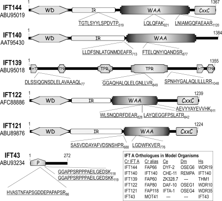
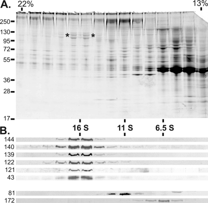
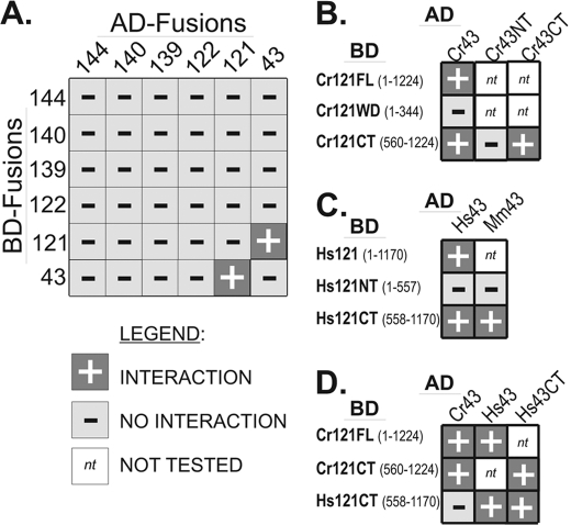
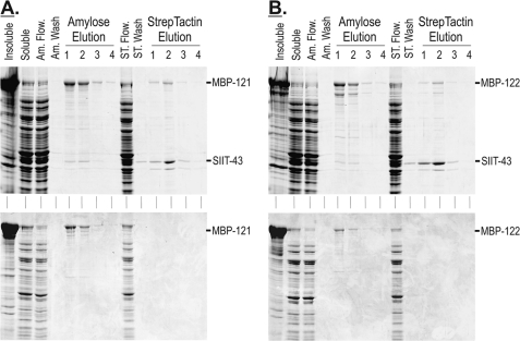
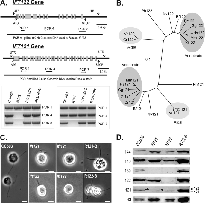
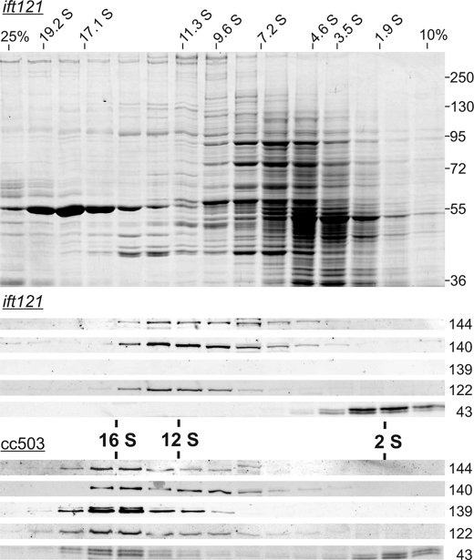
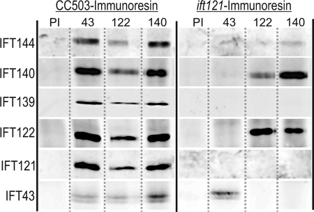
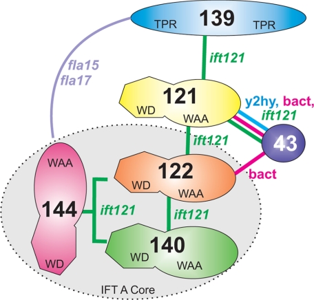
Chlamydomonas reinhardtii intraflagellar transport (IFT) particles can be biochemically resolved into two smaller assemblies, complexes A and B, that contain up to six and 15 protein subunits, respectively. We provide here the proteomic and immunological analyses that verify the identity of all six Chlamydomonas A proteins. Using sucrose density gradient centrifugation and antibody pulldowns, we show that all six A subunits are associated in a 16 S complex in both the cell bodies and flagella. A significant fraction of the cell body IFT43, however, exhibits a much slower sedimentation of ∼2 S and is not associated with the IFT A complex. To identify interactions between the six A proteins, we combined exhaustive yeast-based two-hybrid analysis, heterologous recombinant protein expression in Escherichia coli, and analysis of the newly identified complex A mutants, ift121 and ift122. We show that IFT121 and IFT43 interact directly and provide evidence for additional interactions between IFT121 and IFT139, IFT121 and IFT122, IFT140 and IFT122, and IFT140 and IFT144. The mutant analysis further allows us to propose that a subset of complex A proteins, IFT144/140/122, can form a stable 12 S subcomplex that we refer to as the IFT A core. Based on these results, we propose a model for the spatial arrangement of the six IFT A components.


Figures

FIGURE 1.
FIGURE 1.
Chlamydomonas IFT A protein analysis. Sucrose density gradient-purified IFT A proteins were separated by electrophoresis with isolated tryptic peptides analyzed by Edman degradation as described under “Experimental Procedures.” IFT43 was excised from an SDS-PAGE gel of immunopurified IFT A (see supplemental Fig. S1) prior to trypsinization and MALDI-TOF mass spectroscopy. Three peptide masses resulting from the IFT43 analysis, 2122.9794, 1874.9612, and 2003.0562 Da, matched predicted masses of three peptide sequences, HVASTNFAPSGDDEPAPAPSR86, GGAPPSRPPPAEILGEDSK118, and GGAPPSRPPPAEILGEDSKK119, respectively, that came from a single ORF in the Chlamydomonas genome. The third IFT43 peptide resulted from an incomplete cleavage at Lys119. WD, WD repeat domain; IR, intervening region; WAA, degenerate TPR-like repeats; CXXC, Cys-Xaa-Xaa-Cys repeat domain; TPR, tetratricopeptide repeats; TPRV, TPR domain present in vertebrate THM1/IFT139; HT, half of a tetratricopeptide repeat; P, proline-rich domain.
FIGURE 2.
FIGURE 2.
The IFT A complex is stable to increased ionic strength. The membrane plus matrix proteins were extracted from the flagella isolated from 16 liters of fla2 cells and fractionated through a 13-ml 10–25% sucrose density gradient (SW41Ti) in HMDEK buffer containing 300 mm NaCl. A, fractions 7–21 (21.5–13% sucrose) were resolved on a 7.5–15% acrylamide SDS-PAGE gel and stained with Coomassie Blue; the protein streaking visible in the upper half of the gel was due to the high salt present in the gradient fractions. The asterisks (*) denote the higher molecular mass IFT A subunits found at ∼16 S. Sucrose concentrations are shown at the top, whereas the positions of protein mass standards are indicated on the left. B, immunoblot analysis of fractions 7–21. Antibodies to specific IFT subunits are labeled on the left. All six of the IFT A subunits co-sediment at ∼16 S under these conditions. The anti-IFT81 (81.1) indicates the ∼11 S location of the IFT complex B core; the 6.5 S peak of IFT172 (172.1) serves as an example of a peripheral B component that dissociates from the B core in the presence of 300 mm NaCl.
FIGURE 3.
FIGURE 3.
Directed yeast-based two-hybrid analysis reveals an IFT121-IFT43 interaction. IFT A proteins were fused with either the AD or BD of the GAL4 transcriptional activator and assayed for interaction in yeast. A, exhaustive two-hybrid analysis was carried out using all six of the Chlamydomonas IFT A proteins. All possible combinations were tested by mating a library of AH109 yeast cells with a library of Y187 cells. The AH109 library represented a mixture of cells that contained all six AD-IFT A fusions, whereas the Y187 library represented a mixture of cells that contained all six of the BD-IFT A fusions. Using this approach, only IFT43 and IFT121 showed an interaction. B, testing for interactions between the Chlamydomonas IFT43 and portions of IFT121 revealed that the N-terminal WD40 domain of IFT121 was neither sufficient nor required for the interaction with IFT43. C, the human and murine IFT43 interacted with the C-terminal half but not the N-terminal half of human IFT121. D, the Chlamydomonas IFT43 failed to interact with the C-terminal half of human IFT121 but human IFT43 did interact with both the human and algal IFT121. Interactions are denoted by +; no interaction is denoted by −; not tested is denoted by nt.
FIGURE 4.
FIGURE 4.
Recombinant IFT43 co-purifies with recombinant IFT121 and IFT122. A, upper panel, parallel purification of co-expressed MBP-121 and SIIT-43 using amylose and StrepTactin chromatographies. A, lower panel, control expression of MBP-121 shows that MBP-121 did not bind to and elute from the StrepTactin resin when expressed in the absence of SIIT-IFT43. B, upper panel, parallel purification of co-expressed MBP-122 and SIIT-43 using amylose and StrepTactin chromatographies. B, lower panel, control expression of MBP-122 shows that MBP-122 did not bind to and elute from the StrepTactin resin when expressed in the absence of SIIT-43. Insoluble, insoluble bacterial proteins; soluble, soluble extract (column load); Am. Flow., amylose resin flow-through; Am. Wash, amylose resin final wash; ST Flow., StrepTactin resin flow-through; ST Wash, StrepTactin resin final wash.
FIGURE 5.
FIGURE 5.
The loss of Chlamydomonas IFT121 or IFT122 results in distinctive disruptions to IFT complex A, both of which severely disrupt ciliogenesis. A, genomic maps and PCR analysis of genomic DNA isolated from wild-type, mutant, and rescued strains. R122-BP1, R122-BP2, and R121-BP1 represent mutant strains that were rescued with PCR-amplified genomic DNA, whereas R121-BAC represents an ift121 mutant strain that was rescued using intact BAC DNA. B, taxonomic relatedness of the IFT122 and IFT121 gene products. Cr, C. reinhardtii; Vc, Volvox carteri; Hs, Homo sapiens; Mm, Mus musculus; Gg, Gallus gallus; Xt, Xenopus tropicalis; Dr, Danio rerio; Bf, Branchiostoma floridae; Nv, Nematostella vectensis; Ph, Pediculus humanus corporis. C, phase-contrast images of ift121 and ift122 strains reveal cells with short or no flagella; >95% of ift121 and ift122 rescued with isolated BAC DNA containing IFT121 or IFT122 or PCR-amplified genomic fragments of the IFT121 and IFT122 genes, respectively, recovered assembly of full-length flagella. D, the levels of IFT A proteins extracted from 2 × 10⁶ cells are measured using immunoblots. Soluble protein extracts from whole Chlamydomonas cells were resolved by SDS-PAGE on 10% polyacrylamide gels and transferred to nitrocellulose membrane before probing with antibodies against each of the six IFT A proteins. R122-B represents an ift122 mutant strain in which flagellar assembly was rescued using intact BAC DNA; R121-B represents an ift121 mutant strain rescued using PCR-amplified genomic IFT121.
FIGURE 6.
FIGURE 6.
Co-sedimentation of an IFT A subcomplex extracted from ift121 cells. Whole cell extracts were fractionated on 5.0-ml 10–25% sucrose density gradients. Top, Coomassie-stained 8% polyacrylamide SDS-PAGE gel of ift121 fractionation with sedimentation standard S values shown ranging from 1.9 to 19.2 S. For reference, the prominent ∼55 kDa protein sedimenting at ∼19 S is the large subunit of RuBisCo. Middle, Western blots corresponding to the ift121 fractionation were probed with anti-IFT A antibodies. IFT144, IFT140, and IFT122 co-sediment with a peak of ∼12 S, whereas IFT43 peaks at ∼2 S; IFT121 (not shown) and IFT139 were absent from the extract. Bottom, corresponding Western blots of a sucrose density gradient fractionated whole cell extract from the parental strain, CC-503. Each of the IFT A subunits (IFT121 is not shown) co-sediment at 16–17 S; a fraction of IFT43, however, sedimented at ∼2 S.
FIGURE 7.
FIGURE 7.
Antibody pull-downs of IFT A reveal formation of a subcomplex in ift121. Aliquots of soluble protein from whole cell extracts of CC-503 or ift121 were incubated with preimmune (PI) or subunit-specific antibody resins as indicated above each column. Immunoadsorbed proteins were resolved by SDS-PAGE and transferred to nitrocellulose and probed with anti-IFT A antibodies indicated to the left of each row.
FIGURE 8.
FIGURE 8.
Interaction model of interactions within the Chlamydomonas IFT A complex. The model shown summarizes the IFT A subunit interactions that have been directly visualized, such as IFT121-IFT43, or implied, such as IFT139-IFT121. Based on biochemical analysis of the ift121 mutant, we propose that IFT122, IFT140, and IFT144 assemble to create a heterotrimeric stable core complex. The interactions of IFT121 with both IFT139 and IFT43 are supported by the observation that loss of IFT121 completely removes IFT139 and IFT43 from complex A. Last, IFT139 and IFT144 demonstrate a complex genetic interaction that suggests a functional association (72).

Similar articles

Cited by

References

    1. Afzelius B. A. (1995) Role of cilia in human health. Cell Motil. Cytoskeleton. 32, 95–97 - PubMed
    1. Vincensini L., Blisnick T., Bastin P. (2011) 1001 model organisms to study cilia and flagella. Biol. Cell 103, 109–130 - PubMed
    1. Corbit K. C., Aanstad P., Singla V., Norman A. R., Stainier D. Y., Reiter J. F. (2005) Vertebrate Smoothened functions at the primary cilium. Nature 437, 1018–1021 - PubMed
    1. Eggenschwiler J. T., Anderson K. V. (2007) Cilia and developmental signaling. Annu. Rev. Cell Dev. Biol. 23, 345–373 - PMC - PubMed
    1. Berbari N. F., O'Connor A. K., Haycraft C. J., Yoder B. K. (2009) The primary cilium as a complex signaling center. Curr. Biol. 19, R526–535 - PMC - PubMed

Publication types

MeSH terms