. 2012 Jul;35(3):351-8.
doi: 10.1007/s10143-011-0369-7. Epub 2011 Dec 16.

The role of neuronavigation in intracranial endoscopic procedures

Affiliations

The role of neuronavigation in intracranial endoscopic procedures

Veit Rohde et al. Neurosurg Rev. 2012 Jul.

Abstract

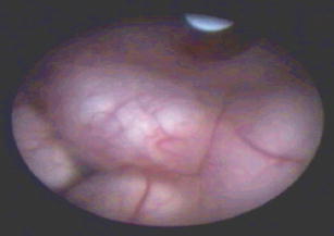
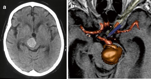
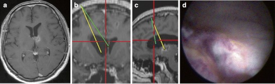
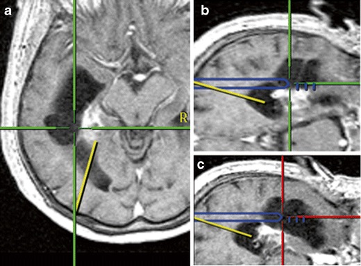
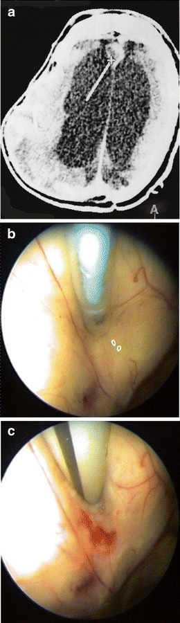
In occlusive hydrocephalus, cysts and some ventricular tumours, neuroendoscopy has replaced shunt operations and microsurgery. There is an ongoing discussion if neuronavigation should routinely accompany neuroendoscopy or if its use should be limited to selected cases. In this prospective clinical series, the role of neuronavigation during intracranial endoscopic procedures was investigated. In 126 consecutive endoscopic procedures (endoscopic third ventriculostomy, ETV, n = 65; tumour biopsy/resection, n = 36; non-tumourous cyst fenestration, n = 23; abscess aspiration and hematoma removal, n = 1 each), performed in 121 patients, neuronavigation was made available. After operation and videotape review, the surgeon had to categorize the role of neuronavigation: not beneficial; beneficial, but not essential; essential. Overall, neuronavigation was of value in more than 50% of the operations, but its value depended on the type of the procedure. Neuronavigation was beneficial, but not essential in 16 ETVs (24.6%), 19 tumour biopsies/resections (52.7%) and 14 cyst fenestrations (60.9%). Neuronavigation was essential in 1 ETV (2%), 11 tumour biopsies/resections (30.6%) and 8 cyst fenestrations (34.8%). Neuronavigation was not needed/not used in 48 ETVs (73.9%), 6 endoscopic tumour operations (16.7%) and 1 cyst fenestration (4.3%). For ETV, neuronavigation mostly is not required. In the majority of the remaining endoscopic procedures, however, neuronavigation is at least beneficial. This finding suggests integrating neuronavigation into the operative routine in endoscopic tumour operations and cyst fenestrations.


Figures

Fig. 1
Fig. 1
Endoscopic view of the third ventricular floor in a patient with occlusive hydrocephalus. The mamillary bodies are clearly visible. The bulging, thick and opaque third ventricular floor does not allow the localization of the basilar artery prior to puncture. Neuronavigation was helpful to select a puncture site in a safe distance to the basilar artery
Fig. 2
Fig. 2
Computerized tomography scan (left) and intraoperative screen display of the navigated endoscope (right) in a 67-year-old female patient with obstructive hydrocephalus due to a giant non-ruptured basilar tip aneurysm. Neuronavigation was essential for opening the third ventricular floor in close vicinity to the basilar tip aneurysm
Fig. 3
Fig. 3
a Magnetic resonance images of a 45-year-old male with a malignant tumour in the caudate nucleus and a monoventricular hydrocephalus. Tumour biopsy and restoration of the CSF flow by septostomy were the aims of the endoscopic procedure. b Intraoperative endoscopic view of the biopsy site. Neuronavigationally defined trajectories for septostomy (c) and tumour biopsy (d). Neuronavigation was considered to be beneficial for septostomy
Fig. 4
Fig. 4
A 70-year-old female with a left temporal AVM which was treated radiosurgically. The patient developed an intraventricular cyst which was progressive in volume and caused a visual field defect. During the endoscopic procedure with neuronavigationally tracked endoscope tip, the small occipital horn was first entered following the predefined yellow trajectory (left), then, on a new trajectory, the cyst was entered (middle) and finally opened to the temporal horn (right). Navigation was essential for ventricle puncture and anatomical orientation
Fig. 5
Fig. 5
Computerized tomography scan axial with the neuronavigationally displayed tip of the endoscope (a) in a 25-year-old patient with a post-traumatic abscess in the interhemispheric fissure. Endoscopically, the anteromedial ventricular wall was perforated (b), and the abscess was aspirated (c). For puncture of the small abscess, neuronavigation was required

References

    1. Ellenbogen RG, Moores LE. Endoscopic management of a pineal and suprasellar germinoma with associated hydrocephalus: technical case report. Minim Invas Neurosurg. 1997;40:13–16. doi: 10.1055/s-2008-1053406. - DOI - PubMed
    1. Grant JA, McLone DG. Third ventriculostomy: a review. Surg Neurol. 1997;47:210–212. doi: 10.1016/S0090-3019(96)00457-0. - DOI - PubMed
    1. Gumbrecht H, Trost HA, Lumenta CB. Neuroendoscopy combined with frameless neuronavigation. Br J Neurosurg. 2000;14:129–131. doi: 10.1080/02688690050004552. - DOI - PubMed
    1. Hopf NJ, Grunert P, Fries G, Resch K, Perneczky A. Endoscopic third ventriculostomy: outcome analysis of 100 consecutive procedures. Neurosurgery. 1999;44:795–806. doi: 10.1097/00006123-199904000-00062. - DOI - PubMed
    1. Jones RFC, Stening WA, Brydon M. Endoscopic third ventriculostomy. Neurosurgery. 1990;26:86–92. doi: 10.1227/00006123-199001000-00012. - DOI - PubMed

MeSH terms

LinkOut - more resources