. 2012 Dec;19(12):1187-95.
doi: 10.1038/gt.2011.197. Epub 2011 Dec 15.

Bacterial magnetic particles as a novel and efficient gene vaccine delivery system

Affiliations
Free PMC article

Bacterial magnetic particles as a novel and efficient gene vaccine delivery system

Y-S Tang et al. Gene Ther. 2012 Dec.
Free PMC article

Abstract

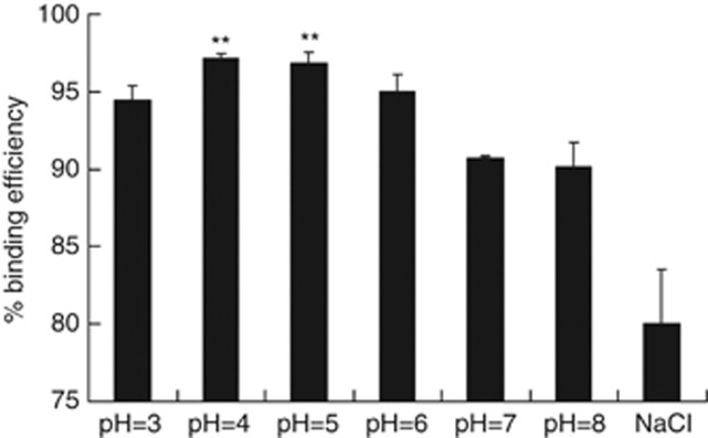
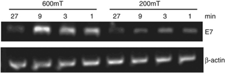
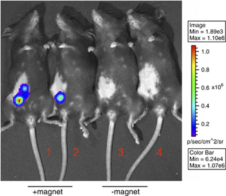
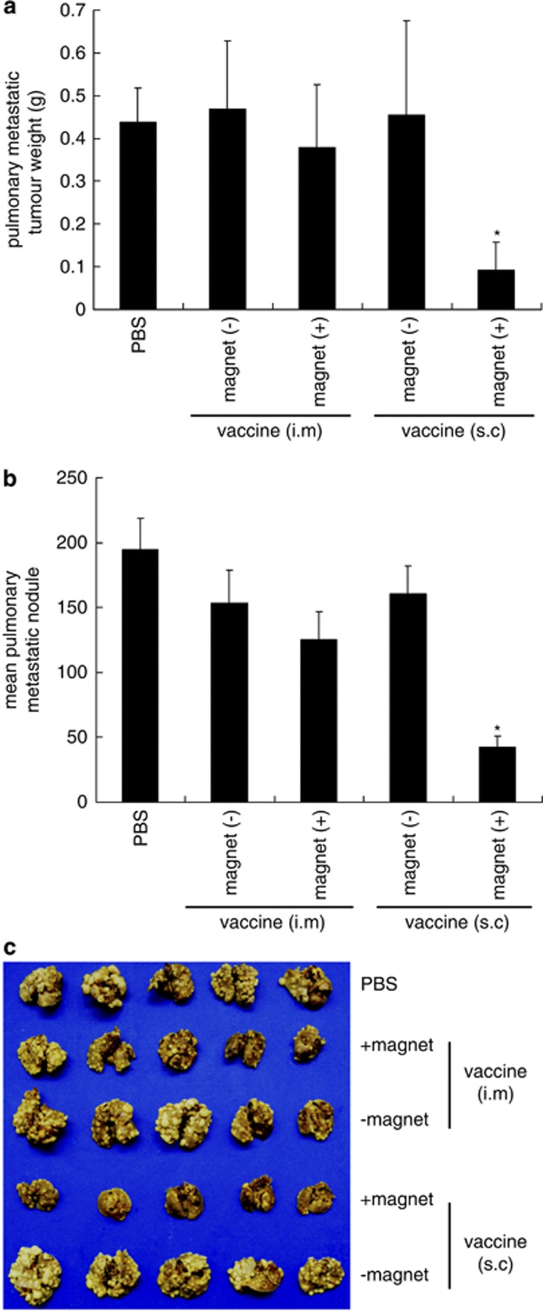
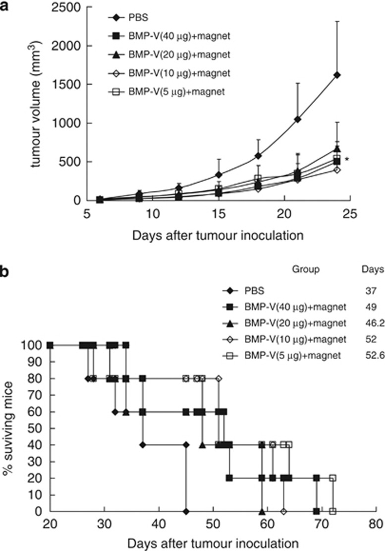
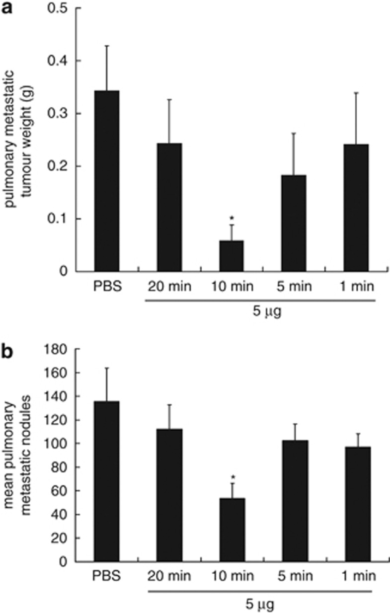
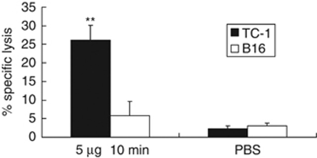
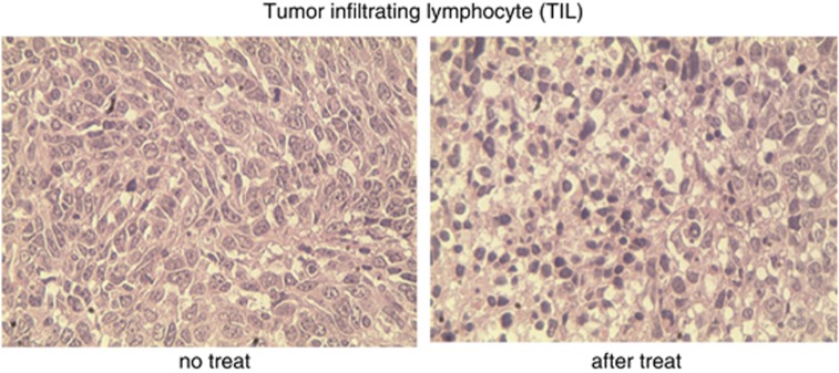
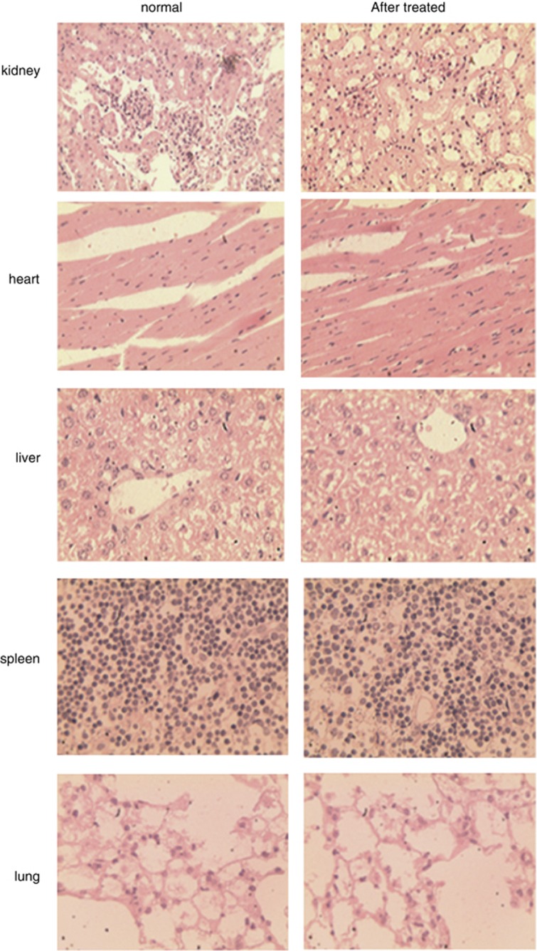
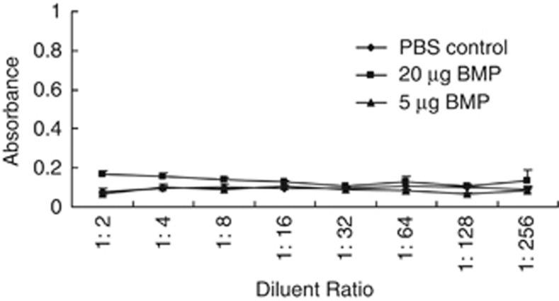
DNA vaccination is an attractive approach for eliciting antigen-specific immunity. In this study, we used magnetosomes (bacterial magnetic particles, BMPs) as carriers of a recombinant DNA composed of a secondary lymphoid tissue chemokine, human papillomavirus type E7 (HPV-E7) and Ig-Fc fragment (pSLC-E7-Fc) to generate a gene vaccine (BMP-V) for tumour immunotherapy. The results indicate that BMPs linked to DNA more efficiently in phosphate-buffered saline (pH=4-5) than in physiological saline. Efficient transfection of BMP-V in vitro and in vivo was achieved when a 600-mT static magnetic field was applied for 10 min. In a mouse tumour model, subcutaneous injection of BMP-V (5 μg, × 3 at 4-day intervals) plus magnetic exposure elicited systemic HPV-E7-specific immunity leading to significant tumour inhibition. The treated mice tolerated BMP-V immunisation well with no toxic side effects, as shown by histopathological examinations of major internal organs. Taken together, these results suggest that BMP can be used as a gene carrier to elicit a systemic immune response.


Figures

Figure 1
Figure 1
The DNA-binding efficiency of BMPs in phosphate-buffered saline at different pH values (pH=3, 4, 5, 6, 7, 8) and 150 mM NaCl. Maximal DNA-binding efficiency was obtained at a pH of 4 and 5. **P<0.05 compared with the 150 mM NaCl group (pH 7).
Figure 2
Figure 2
(a) Bacterial magnetic particles were obtained from Magnetospirillum gryphiswaldense MSR-1. The size distribution of BMPs ranged from 45 to 55 nm. (b) TEM image of BMP-V complexes. Amplification: × 60 000.
Figure 3
Figure 3
Semi-quantitative RT-PCR analysis of B16F10 cells transfected with BMP-V using various magnets and time periods. The expression was measured 48 h after transfection.
Figure 4
Figure 4
Luciferase gene delivery by BMPs-pGL4.17 with or without a magnet. C57BL/6 mice were vaccinated with 10 μg BMPs-pGL4.17 by subcutaneous injection with a magnet on the injection site for 10 min (1, 2) or without a magnet (3, 4). Luciferase imaging was performed at 48 h after vaccination.
Figure 5
Figure 5
Pulmonary tumour nodules and pulmonary metastatic tumour weight in mice vaccinated with BMP-V vaccine through various routes of administration. Mice (n=5) were challenged with TC-1 cells through the tail vein. On day 4, mice were randomised and treated with 20 μg BMP-V in 0.1 ml PBS or PBS alone. The same treatments were repeated on days 8 and 12. All of the mice were killed on day 25. (a) Pulmonary metastatic tumour weights were measured. *P<0.05 compared with the PBS group. (b) Data are presented as the mean number of pulmonary tumour nodules±s.d. of five mice per group in a representative experiment. *Indicates P<0.05 compared with the PBS group. (c) Gross picture of pulmonary tumours in each vaccinated group. Experiments were repeated in duplicate with similar results.
Figure 6
Figure 6
The anti-tumour effects of different amounts of BMP-V. C57BL/6 mice inoculated s.c. with 5 × 10⁴ TC-1 cells. When tumours reached an average diameter of ∼5 mm on day 4, mice were randomised and treated with different doses of BMP-V (40, 20, 10, 5 μg of BMP-V in 0.1 ml PBS or PBS alone). The same treatments were repeated on days 8 and 12. BMP-V were delivered to mice in the shaved inguinal region through subcutaneous injection with a magnet (magnetic flux density of 600 mT) on the injection site for 10 min. (a) Tumour volumes were monitored for 25 days until the control mice began to die. Data are presented as mean tumour volumes±s.d. of five mice per group in a representative experiment. *Indicates P<0.05, all treatments compared with PBS. (b) Survival of mice per treatment group. The results of both experiments were pooled in a stratified analysis, resulting in P>0.05 when the 5 μg BMP-V-treated group was compared with the 10 μg BMP-V-treated group and P<0.05 when the 5 μg BMP-V-treated group was compared with the PBS group. Data are representative of three independent experiments. MST: mean survival time.
Figure 7
Figure 7
Pulmonary tumour nodules and pulmonary metastatic tumour weights in mice vaccinated with BMP-V using various times. Mice (n=5) were challenged with TC-1 cells through the tail vein. On day 4, mice were randomised and treated with 5 μg of BMP-V in 0.1 ml of PBS or PBS alone. The same treatments were repeated on days 8 and 12. BMP-V was administered to mice in the shaved inguinal region through subcutaneous injection with a magnet (magnetic flux density of 600 mT) on the injection site for various times (1, 5, 10, 20 min). All of the mice were killed on day 25. (a) Pulmonary metastatic tumour weights were measured. *P<0.05 compared with the PBS group. (b) Data are presented as the mean number of pulmonary tumour nodules±s.d. of five mice per group in a representative experiment. *Indicates P<0.05 compared with the PBS group.
Figure 8
Figure 8
CTL responses in mice (n=3) immunised with 5 μg of BMP-V plus magnet for 10 min or PBS on days 4, 8 and 12 after TC-1 cell challenge (day 0). On day 18, pooled spleen cells were harvested, and a standard LDH assay was performed. Effector cells from each group were harvested and cytotoxicity was tested against B16 and TC-1 tumour cells at an E:T ratio of 60:1. Data are presented as mean percentage specific lysis±s.d. of triplicate wells of a pooled sample per group in a representative experiment. Experiments were repeated in triplicate with similar results. **Indicates P<0.05, compared with the no-treatment group.
Figure 9
Figure 9
Here, 5 × 10⁴ TC-1 cells were inoculated in the right flank of C57BL/6 mice (n=3) s.c. (day 0). On day 4, mice were randomised and treated with 5 μg BMP-V in 0.1 ml of PBS or PBS alone. The same treatments were repeated on days 8 and 12. BMP-V was administered to mice in the shaved inguinal region through subcutaneous injection with a magnet (magnetic flux density of 600 mT) on the injection site for 10 min. Tumours were excised 3 days after the third vaccination. We performed HE staining of TIL from C57BL/6 mice in the PBS group (left) and the BMP-V treatment group (right). Images are representative of multiple microscopic fields observed in at least three mice per group.
Figure 10
Figure 10
C57BL/6 (n=3) mice inoculated s.c. with 5 × 10⁴ TC-1 cells. On day 4, mice were randomised and treated with 5 μg BMP-V in 0.1 ml PBS. The same treatments were repeated on days 8 and 12. BMP-V was administered to mice in the shaved inguinal region through subcutaneous injection with a magnet (magnetic flux density of 600 mT) on the injection site for 10 min. All of the mice were killed on day 25. We performed HE staining of the kidney, heart, liver, spleen and lung from C57BL/6 mice; normal mice (left), mice treated with BMP-V (right).
Figure 11
Figure 11
BMPs lack immunogenicity. BMPs (20 or 5 μg) were delivered to mice (n=3) in the shaved inguinal region through subcutaneous injection with a magnet (magnetic flux density of 600 mT) on the injection site for 10 min and then were boosted 22 days later. As a negative control, mice were treated with PBS. Seven days after the booster, blood was individually collected from the orbital vein, and the serum was prepared. The serum level of anti-BMP antibodies was determined by ELISA.

Similar articles

Cited by

References

    1. Bansal A, Jackson B, West K, Wang S, Lu S, Kennedy JS, et al. Multifunctional T-cell characteristics induced by a polyvalent DNA prime/protein boost human immunodeficiency virus type 1 vaccine regimen given to healthy adults are dependent on the route and dose of administration. J Virol. 2008;8:6458–6469. - PMC - PubMed
    1. Donnelly J, Berry K, Ulmer JB. Technical and regulatory hurdles for DNA vaccines. Int J Parasitol. 2003;33:457–467. - PubMed
    1. Nicolas JF, Guy B. Intradermal, epidermal and transcutaneous vaccination: from immunology to clinical practice. Expert Rev Vaccines. 2008;7:1201–1214. - PubMed
    1. Bazylinski DA, Frankel RB. Magnetosome formation in prokaryotes. Nat Rev Microbiol. 2004;2:217–230. - PubMed
    1. Bazylinski DA, Garratt-Reed AR, Frankel B. Electron-microscopic studies of magnetosomes in magnetotactic bacteria. Microsc Res Techniq. 1994;27:389–401. - PubMed

Publication types