. 2012 Feb;241(2):270-83.
doi: 10.1002/dvdy.23711. Epub 2011 Dec 14.

Mouse primitive streak forms in situ by initiation of epithelial to mesenchymal transition without migration of a cell population

Affiliations

Mouse primitive streak forms in situ by initiation of epithelial to mesenchymal transition without migration of a cell population

Margot Williams et al. Dev Dyn. 2012 Feb.

Abstract

Background: During gastrulation, an embryo acquires the three primordial germ layers that will give rise to all of the tissues in the body. In amniote embryos, this process occurs via an epithelial to mesenchymal transition (EMT) of epiblast cells at the primitive streak. Although the primitive streak is vital to development, many aspects of how it forms and functions remain poorly understood.

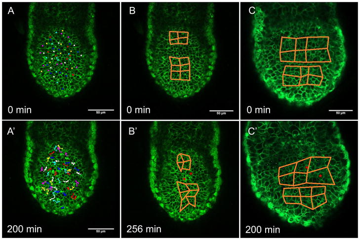
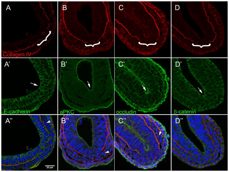
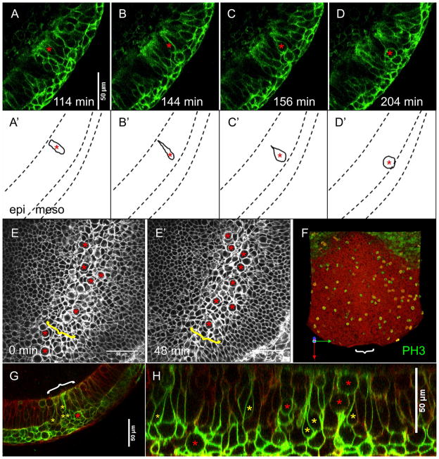
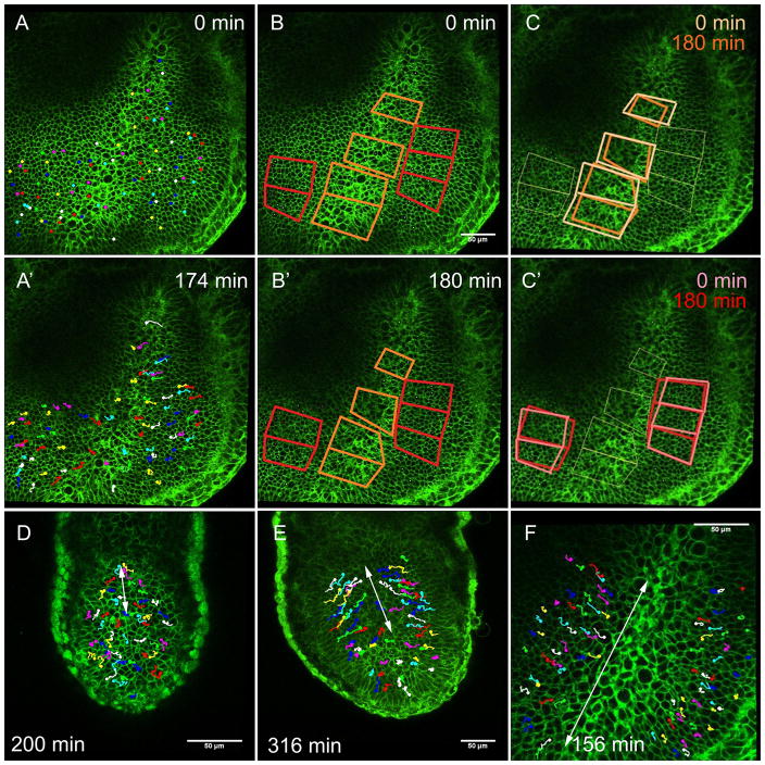
Results: Using live, 4 dimensional imaging and immunohistochemistry, we have shown that the posterior epiblast of the pre-streak murine embryo does not display convergence and extension behavior or large scale migration or rearrangement of a cell population. Instead, the primitive streak develops in situ and elongates by progressive initiation EMT in the posterior epiblast. Loss of basal lamina (BL) is the first step of this EMT, and is strictly correlated with ingression of nascent mesoderm. Once the BL is lost in a given region, cells leave the epiblast by apical constriction in order to enter the primitive streak.

Conclusions: This is the first description of dynamic cell behavior during primitive streak formation in the mouse embryo, and reveals mechanisms that are quite distinct from those observed in other amniote model systems. Unlike chick and rabbit, the murine primitive streak arises in situ by progressive initiation of EMT beginning in the posterior epiblast, without large-scale movement or convergence and extension of epiblast cells.


Figures

Figure 1
Figure 1. The mouse epiblast does not exhibit polonaise movements or convergent extension behavior
Still shots from timelapse movies of live peri-streak EGFP labeled mT/mG pre-streak embryos. A and A′. Cell tracking demonstrates that cells in the posterior epiblast do not undergo bilateral rotational movements, nor do they exhibit anterior displacement. B – C′. Distortion diagrams in the posterior epiblast (posterior identified by nascent mesoderm (red asterisks) in the later timepoints) illustrate that cells of the epiblast do not display convergent extension movements. Distal tip is down.
Figure 2
Figure 2. Primitive streak elongation involves progressive breakdown of basal lamina
A – J. Serial 12 μm transverse sections of mid- to late-streak stage mouse embryos immuno-stained for Laminin (green A–B), Collagen IV (red C–F, green I–K′), and Brachyury (T) (red, I′-K′). Blue is DAPI staining. Posterior is to the right. A – D. Mesodermal cells (red asterisks) are evident in proximal sections of mid-streak stage embryos (A and C), as is a break in the posterior basal lamina (arrows). More distal sections in the same embryos (B and D, respectively) display neither mesoderm nor loss of BL. E – F. In a late-streak stage embryo, loss of basal lamina (arrows) and mesoderm (red asterisks) are evident in proximal (E) and distal (F) sections. G – H. Embryos at early- (G) and late- (H) streak stage. Black lines indicate the approximate level of the indicated transverse sections. Arrowheads indicate distal extent of primitive streak. I – J. Immuno-staining for T confirms the presence of mesoderm (arrows) in proximal sections (I, I′) and absence of mesoderm in distal sections (J, J′) K. Optical sagittal section of a mid-streak stage embryo whole mount immuno-stained for Collagen IV (green) and T (red). Arrows indicate mesoderm cells as confirmed by T staining (K′), arrowhead indicates the distal extent of BL loss.
Figure 3
Figure 3. Loss of basal lamina precedes other steps of EMT within the streak
Transverse sections of late-streak stage embryos. The primitive streak in each embryo (brackets) is identified by a break in Collagen IV staining (red, A–D). Arrows indicate Immuno-fluorescent staining for E-cadherin (green, A′), aPKC (green, B′), Occludin (green, C′), and β-catenin (green, D′) in the epiblast at the site of the primitive streak. Arrowheads indicate lack of staining in the mesoderm layer. Blue in merged images is DAPI. Posterior is to the bottom right.
Figure 4
Figure 4. Epiblast cells enter the primitive streak by apical constriction and somal translocation
A – D. Still shots from a timelapse movie of a live mT/mG:TCre early-bud stage embryo in optical sagittal section. Asterisks mark a cell undergoing apical constriction as it ingresses at the streak. A′ – D′ are representations of still shots A - D to demonstrate changes in the shape of this cell over time. Dashed lines represent borders between germ layers. Epi = epiblast, meso = mesoderm. Mesoderm/ basal surface of epiblast is down. E, E′. Timelapse movie of a live mT/mG no-bud stage embryo viewed en face at the level of the epiblast. The primitive streak (yellow brackets) contains many large, round cells (red asterisks). F. 3 dimensional reconstruction of a whole early head-fold stage embryo immuno-stained for phosphorylated Histone 3 staining (green), as viewed from the inside / epiblast. White bracket marks position of the primitive streak. Distal / anterior is down in E - F. G - H. An optical transverse section (G) and optical sagittal section (H) of a live mT/mG:T Cre no-bud stage embryo show bottle shaped cells (yellow asterisks) and rounded cells (red asterisks) within the primitive streak (white bracket). Mesoderm/ basal surface of epiblast is down.
Figure 5
Figure 5. Cells from the lateral epiblast replenish the primitive streak as cells ingress
A – C′. Still shots from a timelapse movie of a live mT/mG no-bud stage embryo. A. Cell tracking of the posterior epiblast over time (A′). B. Distortion diagrams at the end of the movie (B′) and at the movie’s beginning (B), indicate no convergence and extension. Relationships between cells within the streak are represented by orange diagrams, lateral epiblast by red diagrams. C. Diagrams within the streak are smaller at the end of the movie (dark orange, C) than at the beginning of the movie (light orange, C). Diagrams within the lateral epiblast do not change significantly in size from the movie’s beginning (pink, C′) to its end (red, C′). Distal / anterior is down. D – F. Still shots from timelapse movies of live mT/mG embryos with cell tracking in the epiblast. White arrows indicate regions of medial cell movement in the epiblast. D. In an early-streak stage embryo, cells converge upon the streak only in the posterior third of the epiblast. E. In a mid-streak stage embryo, cells converge upon the streak in the posterior two-thirds of the epiblast. F. In a no-bud stage embryo, epiblast cells converge upon the streak along its entire length. Distal / anterior is down.

References

    1. Acloque H, Ocana OH, Matheu A, Rizzoti K, Wise C, Lovell-Badge R, Nieto MA. Reciprocal Repression between Sox3 and Snail Transcription Factors Defines Embryonic Territories at Gastrulation. Dev Cell. 2011;21:546–558. - PMC - PubMed
    1. Baum B, Settleman J, Quinlan MP. Transitions between epithelial and mesenchymal states in development and disease. Semin Cell Dev Biol. 2008;19:294–308. - PubMed
    1. Bonnerot C, Nicolas JF. Clonal analysis in the intact mouse embryo by intragenic homologous recombination. C R Acad Sci III. 1993;316:1207–1217. - PubMed
    1. Brennan J, Lu CC, Norris DP, Rodriguez TA, Beddington RS, Robertson EJ. Nodal signalling in the epiblast patterns the early mouse embryo. Nature. 2001;411:965–969. - PubMed
    1. Burdsal CA, Damsky CH, Pedersen RA. The role of E-cadherin and integrins in mesoderm differentiation and migration at the mammalian primitive streak. Development. 1993;118:829–844. - PubMed

Publication types