. 2012 Feb;107(4):1157-63.
doi: 10.1152/jn.00401.2011. Epub 2011 Dec 14.

Spatially distinct actions of metabotropic glutamate receptor activation in dorsal lateral geniculate nucleus

Affiliations

Spatially distinct actions of metabotropic glutamate receptor activation in dorsal lateral geniculate nucleus

Gubbi Govindaiah et al. J Neurophysiol. 2012 Feb.

Abstract

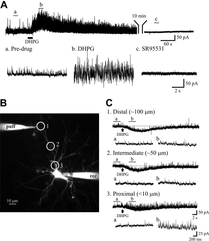
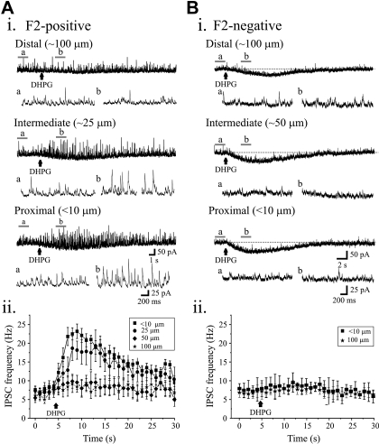
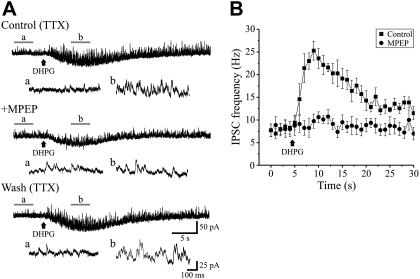
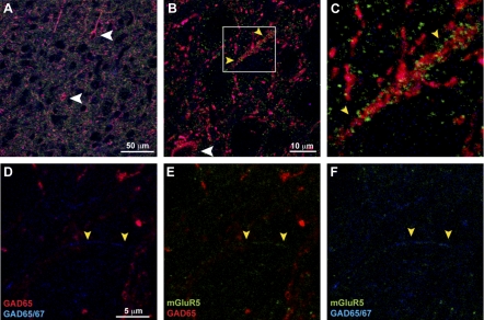
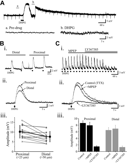
Thalamocortical neurons in the dorsal lateral geniculate nucleus (dLGN) dynamically communicate visual information from the retina to the neocortex, and this process can be modulated via activation of metabotropic glutamate receptors (mGluRs). Neurons within dLGN express different mGluR subtypes associated with distinct afferent synaptic pathways; however, the physiological function of this organization is unclear. We report that the activation of mGluR(5), which are located on presynaptic dendrites of local interneurons, increases GABA output that in turn produces an increased inhibitory activity on proximal but not distal dendrites of dLGN thalamocortical neurons. In contrast, mGluR(1) activation produces strong membrane depolarization in thalamocortical neurons regardless of distal or proximal dendritic locations. These findings provide physiological evidence that mGluR(1) appear to be distributed along the thalamocortical neuron dendrites, whereas mGluR(5)-dependent action occurs on the proximal dendrites/soma of thalamocortical neurons. The differential distribution and activation of mGluR subtypes on interneurons and thalamocortical neurons may serve to shape excitatory synaptic integration and thereby regulate information gating through the thalamus.


Figures

Fig. 1.
Fig. 1.
Proximal but not distal (RS)-3,5-dihydroxyphenylglycine (DHPG) application increases miniature inhibitory synaptic current (mIPSC) frequency in dorsal lateral geniculate nucleus (dLGN) thalamocortical neurons. A: current trace showing mIPSCs recorded from a thalamocortical neuron at holding potential of 0 mV in the presence of TTX (1 μM). The group I metabotropic glutamate receptor (mGluR1) agonist DHPG (25 μM) increases the frequency of mIPSCs (“b”) relative to baseline levels (“a”). The spontaneous IPSC activity is completely attenuated by GABAA in receptor antagonist SR-95531 (“c,” 10 μM). B: typical thalamocortical neuron loaded with Alexa Fluor 594 (50 μM) to perform focal DHPG application under visual guidance. Whole cell recordings were obtained from the soma (rec), whereas DHPG was focally applied at different locations surrounding the neuron as indicated by circles. C: representative current traces obtained from a dLGN thalamocortical neuron in the presence of TTX (1 μM) to show the effects of focal DHPG application at different locations. At distal site (100 μm), DHPG produces only an inward current. At more proximal sites, 50 and <10 μm, DHPG application produces a robust increase in the mIPSC frequency (“b”) compared with baseline activity (“a”) in addition to the inward current. Representative current traces before (“a”) and following DHPG application (“b”) are shown in expanded time scale at the bottom. The expanded traces at the bottom correspond to the underlined region in the top traces.
Fig. 2.
Fig. 2.
Lack of DHPG-mediated increases in mIPSC activity in subsets of dLGN thalamocortical neurons. Ai: current recording from dLGN thalamocortical neuron in presence of TTX (1 μM) reveals strong increase in mIPSC activity following DHPG (100 μM) application at proximal and intermediate sites (F2-positive). DHPG application to distal region did not alter mIPSC activity. Aii: graph illustrates the time course of the DHPG-mediated alterations in mIPSC frequency at different locations. The frequency of mIPSCs were binned for 1 s and shown for 5 s before and 25 s following DHPG application. Bi: representative current traces obtained from a dLGN thalamocortical neuron in presence of TTX (1 μM) reveals no alterations in mIPSC activity following DHPG (100 μM) application (F2-negative). However, DHPG produce inward currents in this neuron. Bii: plot revealing time course of mIPSC frequency over time starting 5 s before and 25 s following DHPG application. The data are plotted as means ± SE.
Fig. 3.
Fig. 3.
Proximal activation of mGluR5 increases mIPSCs in dLGN neurons. A: DHPG (100 μM) application to proximal dendrites increases mIPSC frequency (“b”) relative to baseline (“a”). The increase in mIPSC frequency by DHPG is attenuated by the mGluR5 antagonist 2-methyl-6-(phenylethynyl)pyridine hydrochloride (MPEP; 50 μM) in a reversible manner. The expanded traces at the bottom correspond to the underlined region in the top traces. B: the graph illustrates the time course of the DHPG-mediated alterations in mIPSC frequency. Individual bar represents the number of events in consecutive 1-s bins for 5 s before and 25 s following DHPG application. Values are plotted as means ± SE.
Fig. 4.
Fig. 4.
Expression of mGluR5 and glutamic acid decarboxylase (GAD) in rat dLGN. A and B: colocalization of mGluR5 (green) with GAD65 (red) and GAD65/67 (blue) in cell bodies (white arrowheads) and processes (yellow arrowheads) of GABAergic interneurons. C: higher magnification of region in B illustrating colocalization of mGluR5 in a GAD-containing process. D–F: higher magnification of a representative region showing processes (yellow arrowheads) labeled with GAD67 alone that colocalize with mGluR5 immunoreactivity.
Fig. 5.
Fig. 5.
Activation of postsynaptic mGluR1 but not mGluR5 elicits membrane depolarization in dLGN thalamocortical neurons. A: representative voltage trace from a dLGN neuron revealing that DHPG produces a robust depolarization. Traces are shown in expanded time scale before (“a”) and following exposure to DHPG (“b”). Downward deflections are the hyperpolarizing current pulses (20 pA, 50 ms) to monitor changes in input resistance. Bi: voltage traces from a representative thalamocortical neuron reveals that focal DHPG (50 μM) application to either proximal or distal sites elicits membrane depolarization. DHPG application is indicated by black dots (●). Bii: overlapping of DHPG-mediated responses to proximal and distal sites reveals that the responses to proximal sites is larger than to distal sites. Biii: scatterplot illustrating the amplitude of the DHPG-mediated depolarizations in response to proximal and distal application. Ci: in a different thalamocortical neuron, focal DHPG (50 μM) application elicited consistent membrane depolarization when applied at 30-s intervals. The membrane depolarization is not significantly altered following bath application of MPEP (50 μM); however, the selective mGluR1 antagonist LY 367385 (100 μM) attenuates the DHPG-mediated depolarization. DHPG application is indicated by black dots (●). Cii: average traces for each condition are shown. Ciii: population data revealing that LY 367385 but not MPEP significantly reduces (P < 0.0004, paired t-test) amplitude of membrane depolarization elicited by both proximal and distal DHPG application.

References

    1. Alexander GM, Godwin DW. Presynaptic inhibition of corticothalamic feedback by metabotropic glutamate receptors. J Neurophysiol 94: 163–175, 2005 - PubMed
    1. Alexander GM, Godwin DW. Unique presynaptic and postsynaptic roles of Group II metabotropic glutamate receptors in the modulation of thalamic network activity. Neuroscience 141: 501–513, 2006 - PubMed
    1. Conn PJ, Pin JP. Pharmacology and functions of metabotropic glutamate receptors. Annu Rev Pharmacol Toxicol 37: 205–237, 1997 - PubMed
    1. Cox CL, Sherman SM. Control of dendritic outputs of inhibitory interneurons in the lateral geniculate nucleus. Neuron 27: 597–610, 2000 - PubMed
    1. Cox CL, Zhou Q, Sherman SM. Glutamate locally activates dendritic outputs of thalamic interneurons. Nature 394: 478–482, 1998 - PubMed

Publication types

MeSH terms

LinkOut - more resources