Comparative Study
. 2011 Dec 27;108(52):20976-81.
doi: 10.1073/pnas.1115973108. Epub 2011 Dec 14.

Dextran hydrogel scaffolds enhance angiogenic responses and promote complete skin regeneration during burn wound healing

Affiliations
Comparative Study

Dextran hydrogel scaffolds enhance angiogenic responses and promote complete skin regeneration during burn wound healing

Guoming Sun et al. Proc Natl Acad Sci U S A. .

Abstract

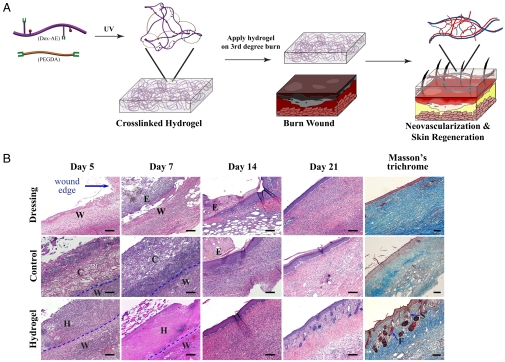
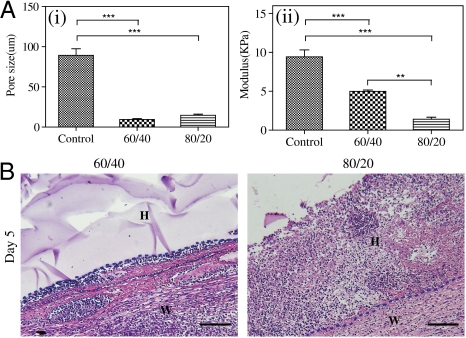
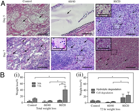
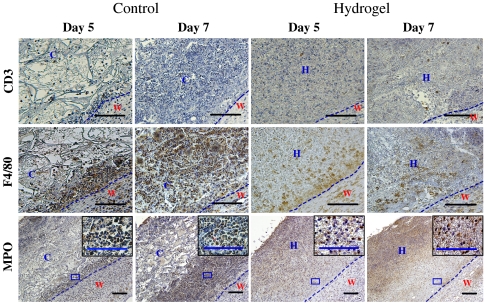
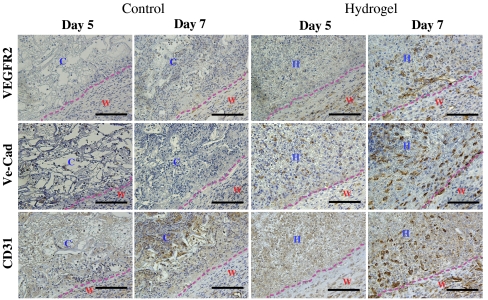
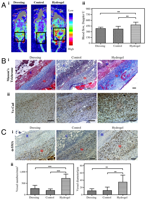
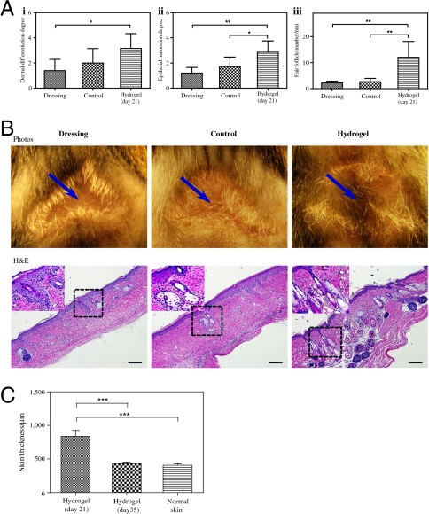
Neovascularization is a critical determinant of wound-healing outcomes for deep burn injuries. We hypothesize that dextran-based hydrogels can serve as instructive scaffolds to promote neovascularization and skin regeneration in third-degree burn wounds. Dextran hydrogels are soft and pliable, offering opportunities to improve the management of burn wound treatment. We first developed a procedure to treat burn wounds on mice with dextran hydrogels. In this procedure, we followed clinical practice of wound excision to remove full-thickness burned skin, and then covered the wound with the dextran hydrogel and a dressing layer. Our procedure allows the hydrogel to remain intact and securely in place during the entire healing period, thus offering opportunities to simplify the management of burn wound treatment. A 3-week comparative study indicated that dextran hydrogel promoted dermal regeneration with complete skin appendages. The hydrogel scaffold facilitated early inflammatory cell infiltration that led to its rapid degradation, promoting the infiltration of angiogenic cells into the healing wounds. Endothelial cells homed into the hydrogel scaffolds to enable neovascularization by day 7, resulting in an increased blood flow significantly greater than treated and untreated controls. By day 21, burn wounds treated with hydrogel developed a mature epithelial structure with hair follicles and sebaceous glands. After 5 weeks of treatment, the hydrogel scaffolds promoted new hair growth and epidermal morphology and thickness similar to normal mouse skin. Collectively, our evidence shows that customized dextran-based hydrogel alone, with no additional growth factors, cytokines, or cells, promoted remarkable neovascularization and skin regeneration and may lead to novel treatments for dermal wounds.


Conflict of interest statement

The authors declare no conflict of interest.

Figures

Fig. 1.
Fig. 1.
Dextran hydrogel as a therapeutic modality. (A) Dextran-based hydrogel promotes neovascularization: Precise structure manipulation allows us to achieve rapid, efficient, and functional neovascularization. (B) Representative images of H&E-stained histological sections at time intervals show that dextran hydrogel promoted wound healing with complete skin appendage regeneration. Masson’s trichrome staining indicates distinct collagen structures formed in dermal layer by day 21. Wound edge indicates the excision rim. W, wound area; H, hydrogel scaffold; C, control scaffold; E, eschar; F, follicle; S, sebaceous gland. Scale bars, 100 μm.
Fig. 2.
Fig. 2.
Characterization of scaffold treatments. Dextran hydrogels (60/40 and 80/20) and cross-linked bovine tendon collagen and glycosaminoglycan scaffolds (control scaffold) were analyzed for (A) (i) porosity and (ii) mechanics. (B). Representative H&E-stained image on day 5 of low ratio (60/40) and high ratio (80/20). W, wound area; H, hydrogel scaffold. Scale bars, 100 μm.
Fig. 3.
Fig. 3.
Hydrogel degradation. (A) Representative images of H&E-stained histological sections of control scaffold (Left), low-ratio dextran hydrogel (Center), and high-ratio dextran hydrogel (Right) on days 5 and 7 of treatment show gel fragmentation (indicated by arrows and magnified inserts). (B) In vitro degradation of control scaffold and hydrogels measured by (i) the total change in scaffold mass and (ii) the relative contribution of HL60 cells and hydrolysis after 72 h. Scale bars, 100 μm (40 μm in insets).
Fig. 4.
Fig. 4.
Inflammatory cell infiltration. Histological sections of control scaffold-treated and hydrogel-treated wounds (Left and Right, respectively) on days 5 and 7 of treatment, stained for CD3 (T cell), F4/80 (macrophage), and MPO (neutrophil). High magnification corresponds to boxed area in the low-magnification images. The dotted line represents the interface between wound and dressing (control scaffold or hydrogel). W, wound area; H, hydrogel scaffold; C, control scaffold. Scale bars, 100 μm.
Fig. 5.
Fig. 5.
Angiogenic cell infiltration. Histological sections of control scaffold-treated and hydrogel-treated wounds (Left and Right, respectively) on days 5 and 7 of treatment, stained for VEGFR2 (Top), VE-Cad (Middle), and CD31 (Bottom). The dotted line represents the interface between wound and control scaffold or hydrogel. W, wound area; H, hydrogel scaffold; C, control scaffold. Scale bars, 100 μm.
Fig. 6.
Fig. 6.
Angiogenic response in day 7. (A) Doppler images of angiogenic response to wound injuries (i), and quantification (ii).The square indicates the wound area under Doppler. (B) Masson’s staining (i) and VE-Cad staining (ii) of wound sites. Collagen layers were formed on the control (untreated) wounds, whereas no such layers formed on control scaffold-treated and hydrogel-treated wounds by day 7; we observed functional blood cells in the hydrogel-treated wounds. (C) Photo of α-SMA staining (i) and quantification based on α-SMA staining (ii) of the wound areas. W, wound area; E, eschar; H, hydrogel scaffold; D, dressing; C, control scaffold. Significance levels were set at: *p < 0.05, **p < 0.01, and ***p < 0.001. Values shown are means ± SD. Scale bars, 100 μm.
Fig. 7.
Fig. 7.
Evaluation of regenerated skin structures. (A) Quantification of skin structures in terms of dermal differentiation (i), epithelial maturation (ii), and the number of hair follicles (iii). (B) A 5-week-long study further demonstrated that dextran hydrogels promote complete skin regeneration with new hair growth, as shown by photos (arrows indicate the center of the original wound; Upper) and H&E-stained histologic sections. High magnification corresponds to boxed area in the low-magnification images. (C) Quantification of skin thickness after 3-week and 5-week-long treatment compared to normal mouse skin. Significance levels were set at: *p < 0.05, **p < 0.01, and ***p < 0.001. Values shown are means ± SD. Scale bars, 100 μm.

References

    1. Zhang X, et al. Association of increasing burn severity in mice with delayed mobilization of circulating angiogenic cells. Arch Surg. 2010;145:259–266. - PMC - PubMed
    1. Sen S, Greenhalgh D, Palmieri T. Review of burn injury research for the year 2009. J Burn Care Res. 2010;31:836–848. - PubMed
    1. Fagenholz PJ, Sheridan RL, Harris NS, Pelletier AJ, Camargo CA., Jr National study of Emergency Department visits for burn injuries, 1993 to 2004. J Burn Care Res. 2007;28:681–690. - PubMed
    1. Zhang X, et al. Impaired angiogenesis and mobilization of circulating angiogenic cells in HIF-1α heterozygous-null mice after burn wounding. Wound Repair Regen. 2010;18:193–201. - PMC - PubMed
    1. Kirker KR, Luo Y, Nielson JH, Shelby J, Prestwich GD. Glycosaminoglycan hydrogel films as bio-interactive dressings for wound healing. Biomaterials. 2002;23:3661–3671. - PubMed

Publication types

MeSH terms

Substances