. 2011 Dec 14;31(50):18198-210.
doi: 10.1523/JNEUROSCI.2560-11.2011.

Neurosteroidogenesis is required for the physiological response to stress: role of neurosteroid-sensitive GABAA receptors

Affiliations

Neurosteroidogenesis is required for the physiological response to stress: role of neurosteroid-sensitive GABAA receptors

Jhimly Sarkar et al. J Neurosci. .

Abstract

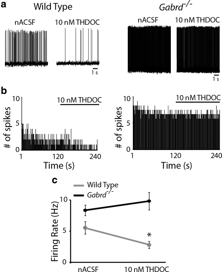
The hypothalamic-pituitary-adrenal (HPA) axis, which mediates the body's response to stress, is largely under GABAergic control. Here we demonstrate that corticotropin-releasing hormone (CRH) neurons are modulated by the stress-derived neurosteroid, tetrahydrodeoxycorticosterone (THDOC), acting on δ subunit-containing GABA(A) receptors (GABA(A)Rs). Under normal conditions, THDOC potentiates the inhibitory effects of GABA on CRH neurons, decreasing the activity of the HPA axis. Counterintuitively, following stress, THDOC activates the HPA axis due to dephosphorylation of KCC2 residue Ser940, resulting in a collapse of the chloride gradient and excitatory GABAergic transmission. The effects of THDOC on CRH neurons are mediated by actions on GABA(A)R δ subunit-containing receptors since these effects are abolished in Gabrd(-/-) mice under both control and stress conditions. Interestingly, blocking neurosteroidogenesis with finasteride is sufficient to block the stress-induced elevations in corticosterone and prevent stress-induced anxiety-like behaviors in mice. These data demonstrate that positive feedback of neurosteroids onto CRH neurons is required to mount the physiological response to stress. Further, GABA(A)R δ subunit-containing receptors and phosphorylation of KCC2 residue Ser940 may be novel targets for control of the stress response, which has therapeutic potential for numerous disorders associated with hyperexcitability of the HPA axis, including Cushing's syndrome, epilepsy, and major depression.


Figures

Figure 1.
Figure 1.
Characterization of CRH-GFP mice. a, Cre recombinase expression in CRH-Cre mice (image adapted from GenSat Brain Atlas). b, LacZ reactivity (black) in the fluorogold-labeled PVN (purple) from offspring of CRH-Cre mice crossed with Rosa26 reporter mice. The magnified image demonstrates the colocalization of LacZ and fluorogold in neurons within the PVN (top right). d, GFP immunoreactivity in CRH-GFP mice generated by crossing CRH-Cre mice with mTomato reporter mice. The magnified image shows the colocalization of GFP (green) and fluorogold (purple) in the CRH-GFP mice (bottom right). Nearly all the LacZ and GFP-positive neurons were also positive for fluorogold (c). In contrast, only ∼50% of fluorogold-positive neurons were also positive for GFP or LacZ (c). n = 6 mice per experimental group. e, Single-cell PCR products from individual CRH-GFP neurons in the PVN run on an ethidium bromide-stained 2% agarose gel. The primers used are shown in Table 1. Lanes 1–10 are the products from individual CRH-GFP neurons. “No primers” lanes are two samples in which the PCR did not contain specific primers. Control cDNA lanes are the product of PCRs using control cDNA as the template (Clontech). f, All of the CRH-GFP-positive neurons tested exhibited a PCR product for both CRH and Gabrd, but not oxytocin (OXT) or thyrotropin-releasing hormone (TRH). n = 10 cells per experimental group.
Figure 2.
Figure 2.
GABAAR δ subunit expression in the PVN. a, A representative Western blot demonstrating the relative abundance of the GABAAR δ subunit in the total protein isolated from the cerebellum, PVN, and hippocampus in wild-type and Gabrd−/− mice. b, The average optical density of GABAAR δ subunit expression in the cerebellum, PVN, and hippocampus from wild-type and Gabrd−/− mice. c, Representative gray scale images of GABAAR δ subunit immunoreactivity in the wild-type PVN and hippocampus compared with the Gabrd−/− PVN. d, The average optical density of GABAAR δ subunit demonstrates expression in the PVN and hippocampus in sections from wild-type mice, but not Gabrd−/− mice. n = 3 mice per experimental group; statistical significance of *p < 0.05 compared with Gabrd−/− levels using a one-way ANOVA with Bonferroni correction for multiple comparisons.
Figure 3.
Figure 3.
Neurosteroid regulation of CRH neurons. a, Representative traces of sIPSCs from GFP-positive CRH neurons in the presence or absence of 10 nm THDOC. b, The superimposed average sIPSCs in the presence or absence of 10 nm THDOC highlight that there is no effect of THDOC on the average decay time (τw) (see inset). c, The average histograms demonstrate that THDOC did not significantly alter the frequency or amplitude of sIPSCs. d, Representative traces of the tonic current in CRH-GFP neurons from wild-type and Gabrd−/− mice held at −70 mV throughout the addition of 10 nm THDOC and saturating concentrations of SR95531. e, The average tonic current is significantly decreased in CRH neurons from Gabrd−/− mice compared with wild-type. THDOC (10 nm) enhanced the tonic GABAergic inhibition in CRH-GFP neurons from wild-type mice, but not Gabrd−/− mice. n = 6–12 mice, 15–32 cells; statistical significance of *p < 0.05 using a paired t test.
Figure 4.
Figure 4.
Neurosteroid regulation of CRH neuronal activity. a, Representative traces of the spontaneous firing rate of CRH-GFP neurons from wild-type and Gabrd−/− mice in nACSF and 10 nm THDOC. b, The binned firing rate (1 s bins) in the same representative CRH neuron over time in the presence of nACSF or 10 nm THDOC (where indicated) from a wild-type (left) and Gabrd−/− mouse (right). c, THDOC significantly decreased the basal firing rate of CRH-GFP neurons from wild-type mice, but not Gabrd−/− mice. n = 12–13 cells, 6 mice per experimental group; significance of *p < 0.05 compared with firing rate in nACSF using a paired t test.
Figure 5.
Figure 5.
Local administration of THDOC into the PVN is sufficient to alter HPA axis activity. a, Timeline of blood sample collection, microinfusion, and corticosterone measurements. b, A representative section demonstrating cresyl violet diffusion into the PVN of wild-type mice following microinfusion of 100 nm THDOC. Dotted line represents the extent of cresyl violet staining from the site of the injection site (needle track; arrow). c, THDOC microinfusion into the PVN significantly decreased serum corticosterone levels within 30 min. Vehicle administration did not alter corticosterone levels. n = 6 mice per experimental group; significance of *p < 0.05 compared with basal corticosterone levels using a paired t test.
Figure 6.
Figure 6.
Neurosteroids exacerbate the corticosterone response to stress. a, Average corticosterone levels in paired samples measured before and after treatment with vehicle, THDOC (20 mg/kg) and finasteride (50 mg/kg). b, The average corticosterone levels in paired samples before and after 30 min restraint stress in wild-type animals treated with vehicle, THDOC, or finasteride. c, Average corticosterone levels measured before and after 30 min restraint stress in vehicle, THDOC, and finasteride-treated Gabrd−/− mice. n = 5–11 mice per experimental group; significance of *p < 0.05 using a one-way ANOVA with Bonferroni correction for multiple comparisons.
Figure 7.
Figure 7.
THDOC increases the activity of CRH neurons following stress. a, Representative traces of the basal firing rate of CRH-GFP neurons recorded under perforated patch, current clamp I = 0 conditions from wild-type and Gabrd−/− mice in nACSF and 10 nm THDOC. b, The average firing rate in paired samples in the presence or absence of THDOC in control wild-type mice or mice subjected to 30 min restraint stress. c, The average firing rate of CRH neurons in control or stress Gabrd−/− mice in the presence or absence of THDOC. n = 11–12 cells, 5–6 mice per experimental group; significance of *p < 0.05 compared with the firing rate in nACSF using a paired t test.
Figure 8.
Figure 8.
Depolarizing and excitatory GABAergic responses following stress. a, Representative, hyperpolarizing sIPSPs recorded in CRH-GFP neurons under perforated patch, current clamp I = 0 conditions from control wild-type mice and depolarizing sIPSPs recorded in slices from mice subjected to acute restraint stress. b, The percentage of cells exhibiting depolarizing sIPSPs from control and stress wild-type mice. n = 10–12 cells, 4 mice per experimental group; significance of *p < 0.05 using an unpaired t test. c, Representative traces of the basal firing rate of CRH-GFP-positive neurons recorded under perforated patch, current clamp I = 0 conditions from control and stress wild-type mice in nACSF and SR95531. d, The average firing rate of CRH neurons from control or stress mice in the presence or absence of SR95531. n = 12 cells, 4 mice per experimental group; significance of *p < 0.05 using a paired t test.
Figure 9.
Figure 9.
Dephosphorylation and downregulation of KCC2 following stress. a, Representative Western blot demonstrating decreased expression of KCC2 (left), phosphorylation of KCC2 residue Ser940 (center), and surface biotinylated KCC2 (right) in two independent PVN samples in control and stress mice. b, The average optical density measurements of KCC2 expression in control mice and mice subjected to 30 min restraint stress. c, The average optical density of phosphorylated KCC2 residue Ser940 in the PVN from control or stress mice. d, The percentage of phosphorylated KCC2 is significantly decreased following stress compared with control. e, A Spearman rank order correlation of corticosterone levels and phosphorylation levels of KCC2 residue Ser940 reveals a significant negative correlation (r = −0.6993). f, The average biotinylated KCC2 expression in the PVN of control and stress wild-type mice. n = 8–10 mice per experimental group; significance of *p < 0.05 compared with control using an unpaired t test.
Figure 10.
Figure 10.
Neurosteroidogenesis is required for stress-induced anxiety-like behavior. a, b, The average time spent (a) and the distance traveled (b) in the open arm of the elevated plus maze in control mice and mice subjected to 30 min restraint stress and treated with vehicle, THDOC, or finasteride. c, The average time spent in the center of the open field in control mice and vehicle, THDOC, or finasteride-treated mice subjected to 30 min restraint stress. d, The average locomotor behavior, assessed by the number of beam breaks, in control mice and vehicle, THDOC, or finasteride-treated mice subjected to 30 min restraint stress. n = 11–12 mice per experimental group; *p < 0.05 compared with control, #p < 0.05 compared with vehicle-treated stress using a one-way ANOVA with Bonferroni correction for multiple comparisons.
Figure 11.
Figure 11.
A model of HPA axis regulation. The activity of the HPA axis is governed by CRH neurons in the PVN. These neurons receive inputs from many different brain regions and are modulated by many different neurotransmitter systems. However, the activity of these neurons is ultimately under robust GABAergic control. We propose a model of HPA axis activation which overrides this robust GABAergic inhibition by dephosphorylating and downregulating KCC2, resulting in a collapse in the chloride gradient, and excitatory actions of GABA. The stress-derived neurosteroid, THDOC, potentiates GABAAR δ subunit-containing receptors, resulting in excitation of CRH neurons which is required to mount the physiological response to stress in a rapid, all-or-none fashion.

Similar articles

Cited by

References

    1. Banke TG, McBain CJ. GABAergic input onto CA3 hippocampal interneurons remains shunting throughout development. J Neurosci. 2006;26:11720–11725. - PMC - PubMed
    1. Barnard EA, Skolnick P, Olsen RW, Mohler H, Sieghart W, Biggio G, Braestrup C, Bateson AN, Langer SZ. International Union of Pharmacology. XV. Subtypes of gamma-aminobutyric acidA receptors: classification on the basis of subunit structure and receptor function. Pharmacol Rev. 1998;50:291–313. - PubMed
    1. Belelli D, Casula A, Ling A, Lambert JJ. The influence of subunit composition on the interaction of neurosteroids with GABA(A) receptors. Neuropharmacology. 2002;43:651–661. - PubMed
    1. Ben-Ari Y. Excitatory actions of gaba during development: the nature of the nurture. Nat Rev Neurosci. 2002;3:728–739. - PubMed
    1. Boehm SL, 2nd, Homanics GE, Blednov YA, Harris RA. delta-Subunit GABAA receptor knockout mice are less sensitive to the actions of 4,5,6,7-tetrahydroisoxazolo-[5,4-c]pyridin-3-ol. Eur J Pharmacol. 2006;541:158–162. - PubMed

Publication types

MeSH terms

LinkOut - more resources