Site occupancy and glycan compositional analysis of two soluble recombinant forms of the attachment glycoprotein of Hendra virus
- PMID: 22171062
- PMCID: PMC3287018
- DOI: 10.1093/glycob/cwr180
Site occupancy and glycan compositional analysis of two soluble recombinant forms of the attachment glycoprotein of Hendra virus
Abstract
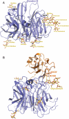
Hendra virus (HeV) continues to cause morbidity and mortality in both humans and horses with a number of sporadic outbreaks. HeV has two structural membrane glycoproteins that mediate the infection of host cells: the attachment (G) and the fusion (F) glycoproteins that are essential for receptor binding and virion-host cell membrane fusion, respectively. N-linked glycosylation of viral envelope proteins are critical post-translation modifications that have been implicated in roles of structural integrity, virus replication and evasion of the host immune response. Deciphering the glycan composition and structure on these glycoproteins may assist in the development of glycan-targeted therapeutic intervention strategies. We examined the site occupancy and glycan composition of recombinant soluble G (sG) glycoproteins expressed in two different mammalian cell systems, transient human embryonic kidney 293 (HEK293) cells and vaccinia virus (VV)-HeLa cells, using a suite of biochemical and biophysical tools: electrophoresis, lectin binding and tandem mass spectrometry. The N-linked glycans of both VV and HEK293-derived sG glycoproteins carried predominantly mono- and disialylated complex-type N-glycans and a smaller population of high mannose-type glycans. All seven consensus sequences for N-linked glycosylation were definitively found to be occupied in the VV-derived protein, whereas only four sites were found and characterized in the HEK293-derived protein. We also report, for the first time, the existence of O-linked glycosylation sites in both proteins. The striking characteristic of both proteins was glycan heterogeneity in both N- and O-linked sites. The structural features of G protein glycosylation were also determined by X-ray crystallography and interactions with the ephrin-B2 receptor are discussed.
Figures


Similar articles
-
Novel Functions of Hendra Virus G N-Glycans and Comparisons to Nipah Virus.J Virol. 2015 Jul;89(14):7235-47. doi: 10.1128/JVI.00773-15. Epub 2015 May 6. J Virol. 2015. PMID: 25948743 Free PMC article.
-
Receptor binding, fusion inhibition, and induction of cross-reactive neutralizing antibodies by a soluble G glycoprotein of Hendra virus.J Virol. 2005 Jun;79(11):6690-702. doi: 10.1128/JVI.79.11.6690-6702.2005. J Virol. 2005. PMID: 15890907 Free PMC article.
-
Recombinant Glycoprotein E of Varicella Zoster Virus Contains Glycan-Peptide Motifs That Modulate B Cell Epitopes into Discrete Immunological Signatures.Int J Mol Sci. 2019 Feb 22;20(4):954. doi: 10.3390/ijms20040954. Int J Mol Sci. 2019. PMID: 30813247 Free PMC article.
-
Function and 3D structure of the N-glycans on glycoproteins.Int J Mol Sci. 2012;13(7):8398-8429. doi: 10.3390/ijms13078398. Epub 2012 Jul 6. Int J Mol Sci. 2012. PMID: 22942711 Free PMC article. Review.
-
Glycan Shielding and Modulation of Hepatitis C Virus Neutralizing Antibodies.Front Immunol. 2018 Apr 27;9:910. doi: 10.3389/fimmu.2018.00910. eCollection 2018. Front Immunol. 2018. PMID: 29755477 Free PMC article. Review.
Cited by
-
Hendra virus and Nipah virus animal vaccines.Vaccine. 2016 Jun 24;34(30):3525-34. doi: 10.1016/j.vaccine.2016.03.075. Epub 2016 May 4. Vaccine. 2016. PMID: 27154393 Free PMC article.
-
A treatment for and vaccine against the deadly Hendra and Nipah viruses.Antiviral Res. 2013 Oct;100(1):8-13. doi: 10.1016/j.antiviral.2013.06.012. Epub 2013 Jul 6. Antiviral Res. 2013. PMID: 23838047 Free PMC article. Review.
-
Henipavirus mediated membrane fusion, virus entry and targeted therapeutics.Viruses. 2012 Feb;4(2):280-308. doi: 10.3390/v4020280. Epub 2012 Feb 13. Viruses. 2012. PMID: 22470837 Free PMC article. Review.
-
Vaccines to Emerging Viruses: Nipah and Hendra.Annu Rev Virol. 2020 Sep 29;7(1):447-473. doi: 10.1146/annurev-virology-021920-113833. Annu Rev Virol. 2020. PMID: 32991264 Free PMC article.
-
Immunization strategies against henipaviruses.Curr Top Microbiol Immunol. 2012;359:197-223. doi: 10.1007/82_2012_213. Curr Top Microbiol Immunol. 2012. PMID: 22481140 Free PMC article. Review.
References
-
- Bishop KA, Stantchev TS, Hickey AC, Khetawat D, Bossart KN, Krasnoperov V, Gill P, Feng YR, Wang L, Eaton BT, et al. Identification of Hendra virus G glycoprotein residues that are critical for receptor binding. J Virol. 2007;81:5893–5901. doi:10.1128/JVI.02022-06. - DOI - PMC - PubMed
-
- Bonaparte MI, Dimitrov AS, Bossart KN, Crameri G, Mungall BA, Bishop KA, Choudhry V, Dimitrov DS, Wang LF, Eaton BT, et al. Ephrin-B2 ligand is a functional receptor for Hendra virus and Nipah virus. Proc Natl Acad Sci USA. 2005;102:10652–10657. doi:10.1073/pnas.0504887102. - DOI - PMC - PubMed
-
- Bossart KN, Broder CC. Paramyxovirus entry. In: Pöhlmann S, Simmons G, editors. Viral Entry into Host Cells. Austin (TX): Landes Bioscience; 2009.
-
- Bossart KN, Crameri G, Dimitrov AS, Mungall BA, Feng YR, Patch JR, Choudhary A, Wang LF, Eaton BT, Broder CC. Receptor binding, fusion inhibition, and induction of cross-reactive neutralizing antibodies by a soluble G glycoprotein of Hendra virus. J Virol. 2005;79:6690–6702. doi:10.1128/JVI.79.11.6690-6702.2005. - DOI - PMC - PubMed
-
- Bowden TA, Aricescu AR, Gilbert RJ, Grimes JM, Jones EY, Stuart DI. Structural basis of Nipah and Hendra virus attachment to their cell-surface receptor ephrin-B2. Nat Struct Mol Biol. 2008;15:567–572. doi:10.1038/nsmb.1435. - DOI - PubMed
Publication types
MeSH terms
Substances
Associated data
- Actions
Grants and funding
LinkOut - more resources
Full Text Sources
Other Literature Sources

