. 2012 Mar 15;21(6):1350-63.
doi: 10.1093/hmg/ddr573. Epub 2011 Dec 13.

Roles of the Drosophila LRRK2 homolog in Rab7-dependent lysosomal positioning

Affiliations

Roles of the Drosophila LRRK2 homolog in Rab7-dependent lysosomal positioning

Mark W Dodson et al. Hum Mol Genet. .

Abstract

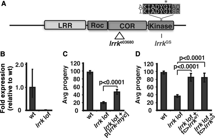
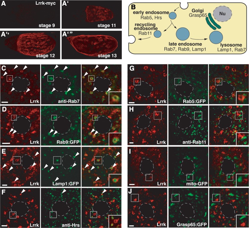
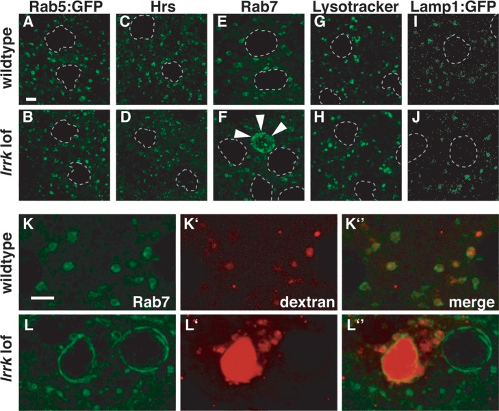
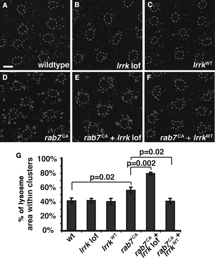
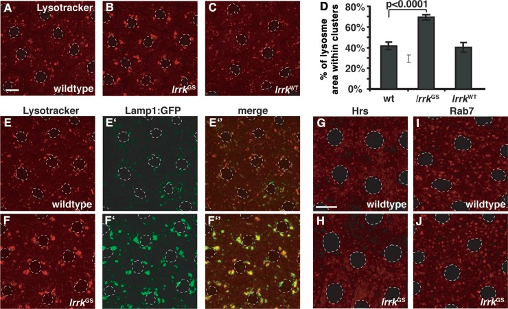
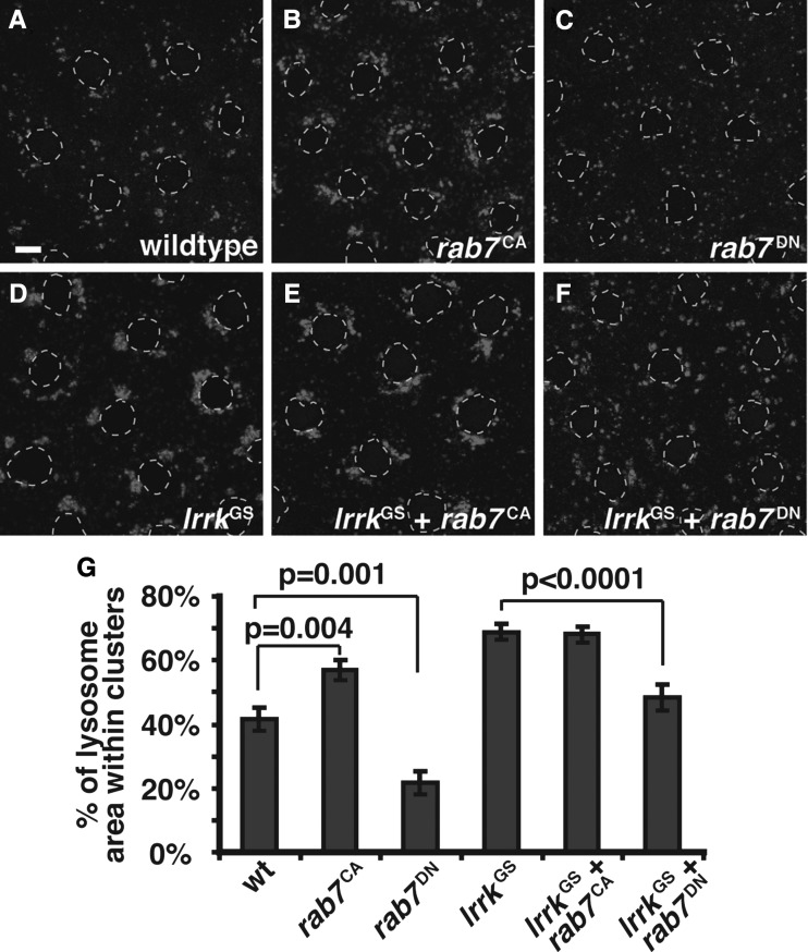
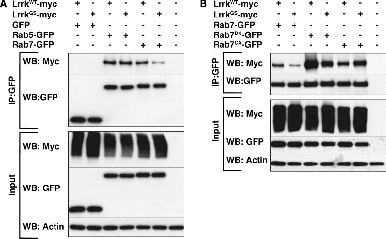
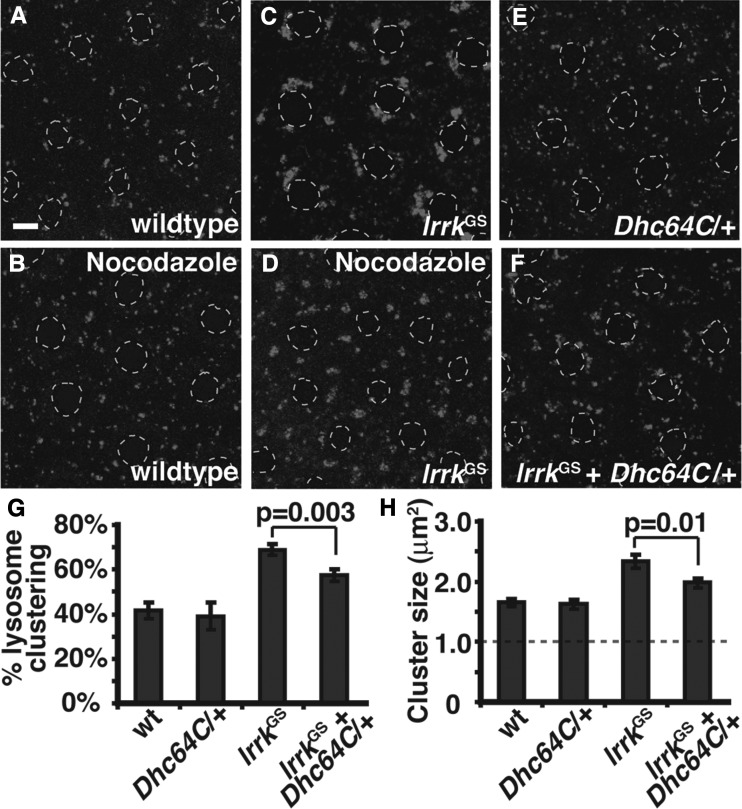
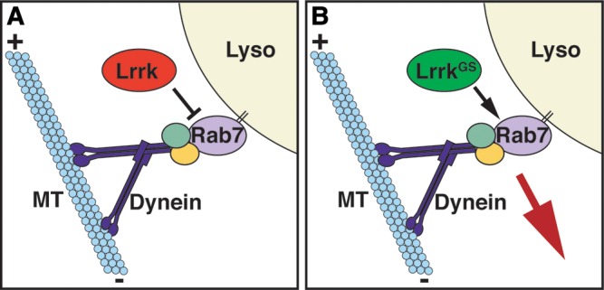
LRRK2 (PARK8) is the most common genetic determinant of Parkinson's disease (PD), with dominant mutations in LRRK2 causing inherited PD and sequence variation at the LRRK2 locus associated with increased risk for sporadic PD. Although LRRK2 has been implicated in diverse cellular processes encompassing almost all cellular compartments, the precise functions of LRRK2 remain unclear. Here, we show that the Drosophila homolog of LRRK2 (Lrrk) localizes to the membranes of late endosomes and lysosomes, physically interacts with the crucial mediator of late endosomal transport Rab7 and negatively regulates rab7-dependent perinuclear localization of lysosomes. We also show that a mutant form of lrrk analogous to the pathogenic LRRK2(G2019S) allele behaves oppositely to wild-type lrrk in that it promotes rather than inhibits rab7-dependent perinuclear lysosome clustering, with these effects of mutant lrrk on lysosome position requiring both microtubules and dynein. These data suggest that LRRK2 normally functions in Rab7-dependent lysosomal positioning, and that this function is disrupted by the most common PD-causing LRRK2 mutation, linking endolysosomal dysfunction to the pathogenesis of LRRK2-mediated PD.


Figures

Figure 1.
Figure 1.
Lrrk is required in follicle cells for proper female fertility. (A) Schematic depicting domain organization of Drosophila Lrrk and location of PiggyBac insertion in the lrrke03680 allele and G-to-S substitution in lrrkGS. Above is an alignment of the amino acid sequences of human LRRK2 (top) and Drosophila Lrrk (bottom) in the region of the GS mutation (marked with an asterisk). LRR, leucine-rich repeats; Roc, GTPase domain; Cor, C-terminal of Roc domain. (B) lrrke03680 homozygous flies produce no detectable lrrk transcript by RT–PCR. (C) Fertility is reduced in lrrke03680 homozygous mutants, and the phenotype is partially rescued by a Myc-tagged genomic rescue transgene (p[lrrk-myc]). (D) Fertility is fully restored to lrrke03680/Df null mutant flies by follicle cell-specific expression of either wild-type lrrk (fc>lrrkWT) or lrrkGS (fc>lrrkGS).
Figure 2.
Figure 2.
Lrrk localizes to late endosomes and lysosomes in follicle cells. (A) Anti-Myc staining of egg chambers from flies carrying a single copy of the C-terminally Myc-tagged lrrk genomic rescue transgene. Lrrk-Myc was not detected in egg chambers prior to stage 11 (A), and persisted through stages 12 (A″) and 13 (A″′). Lrrk-Myc staining reveals a patchy distribution, with expression in subsets of follicle cells (including main body and stretched follicle cells), but not in germline cells. (B) Schematic depicting the endolysosomal pathway and proteins labeling specific compartments. (CJ) Images of single main body follicle cells with Lrrk-Myc staining in red and costaining for various cellular compartments in green. Nuclei are outlined by dashed gray lines, and insets depict enlargements of the boxed regions. Lrrk localizes to discrete cytosolic puncta that often appear as halos surrounding a non-stained central region, indicative of an association with vesicle membranes. These large Lrrk-positive halos colocalize with the late endosomal markers Rab7 (C) and Rab9:GFP (D). The large Lrrk-positive structures also contain Lamp1:GFP (E). Note that while Rab7 and Rab9:GFP label late endosome membranes, Lamp1:GFP accumulates in the lumena of late endosomes and lysosomes. Small Lrrk-positive puncta occasionally colocalize with the early endosomal marker Hrs (F). In contrast, there is little colocalization between Lrrk and Rab5 (G) or Rab11 (H), although these markers often appear adjacent to the Lrrk-positive vesicles. There is no significant colocalization between Lrrk and the mitochondrial marker mitoGFP (I) or the Golgi marker Grasp65:GFP (J). Scale bars represent 5 μm.
Figure 3.
Figure 3.
Altered Rab7-positive late endosomal compartments in lrrk loss-of-function mutants. (A–J) Examination of a battery of endolysosomal markers in wild-type (A, C, E, G, I) and lrrk null mutant (B, D, F, H, J) follicle cells reveals no distinguishable difference between the early endosomal markers Rab5 (A versus B) or Hrs (C versus D), or the lysosomal markers Lysotracker (G versus H) or Lamp1:GFP (I versus J). A small portion of lrrk mutant follicle cells, however, did display dramatically expanded Rab7-positive late endosomes (marked with arrowheads in F) that were never observed in stage-matched wild-type follicle cells (E). (K and L) After a 30 min chase, fluorescently labeled dextran was taken up into wild-type follicle cells and trafficked predominantly to Rab7-positive late endosomes (K). In lrrk mutant follicle cells (L), some enlarged Rab7-positive structures accumulated massive amounts of dextran, while others were devoid of the tracer. Follicle cell nuclei are outlined with dashed gray lines in (A–J). Scale bars represent 5 μm.
Figure 4.
Figure 4.
lrrk and rab7 genetically interact. (A–F) Lysotracker staining of follicle cells from stage 12 egg chambers. In wild-type follicle cells (A), most Lysotracker-positive vesicles are individual and distributed throughout the cytosol, although small clusters are often seen. Neither lrrk loss-of-function (B), nor overexpression of wild-type lrrk (C) significantly alters lysosome distribution. Expression of constitutively active rab7Q67L (D) results in an increase in the formation of Lysotracker-positive clusters, and these tend to localize to the peri-nuclear region. lrrk loss-of-function significantly enhances lysosome clustering in follicle cells expressing rab7Q67L (E), while overexpression of wild-type lrrk significantly reduces rab7Q67L-induced lysosome clustering (F). Follicle cell nuclei are outlined with dashed gray lines in all Lysotracker images. (G) Quantification of lysosome clustering in the genotypes depicted in (A)–(F). Scale bars represent 5 μm.
Figure 5.
Figure 5.
Expression of lrrkGS drives perinuclear positioning and clustering of lysosomes. (A–C) Lysotracker staining of follicle cells from stage 12 egg chambers. In wild-type follicle cells (A), most Lysotracker-positive vesicles are individual and distributed throughout the cytosol, although small clusters are often seen. Expression of lrrkGS (B) in follicle cells causes almost all Lysotracker-positive structures to collapse into one to four compact perinuclear clusters, while equivalent overexpression of lrrkWT (C) results in no significant changes in the distribution of Lysotracker-positive structures relative to the wild-type. (D) Quantification of lysosome clustering from the genotypes depicted in (A)–(C). (E and F) Lysotracker staining of wild-type (E) and lrrkGS-expressing (F) follicle cells also expressing the lysosomal marker Lamp1:GFP. Lamp1:GFP-positive vesicles are clustered in lrrkGS-expressing cells (F′), colocalizing with Lysotracker (F″). Note that Lamp1:GFP accumulates to a much higher degree in lrrkGS-expressing cells (F′) versus wild-type (E′) in images taken with equivalent microscope settings, suggesting that expression of lrrkGS stabilizes Lamp1:GFP. (G–J) Labeling of early endosomes with the marker Hrs (G and H) and late endosomes with the marker Rab7 (I and J) reveals no significant difference between wild-type (G and I) and lrrkGS-expressing cells (H and J), demonstrating that the clustering effect of lrrkGS is specific to lysosomes. Follicle cell nuclei are outlined with dashed gray lines in all images. Scale bar in (A) represents 5 μm in (A)–(F); scale bar in (G) represents 5 μm in (G)–(J).
Figure 6.
Figure 6.
The GS mutation abrogates the ability of lrrk to inhibit rab7-induced lysosome clustering. (A–F) Lysotracker staining of follicle cells from stage 12 egg chambers of the indicated genotypes. Expression of constitutively active Rab7 (rab7Q67L, B) causes perinuclear clustering of Lysotracker-positive structures similar to that seen with expression of lrrkGS (D), while expression of rab7Q67L and lrrkGS together (E) causes a phenotype that is not significantly different from expression of lrrkGS alone. Expression of dominant-negative Rab7 (rab7T22N, C) causes lysosome dispersal. Expression of rab7T22N along with lrrkGS significantly reduces the lrrkGS-induced lysosome clustering. (G) Quantification of lysosome clustering in the indicated genotypes. Follicle cell nuclei are outlined with dashed gray lines in all Lysotracker images. Scale bars represent 5 μm.
Figure 7.
Figure 7.
The GS mutation alters the characteristics of physical binding between Lrrk and Rab7 forms. (A) In lysates from cultured Drosophila S2 cells transfected with the depicted constructs, LrrkWT immunoprecipitates with both Rab7-GFP and Rab5-GFP, but not with GFP alone. The GS mutation reduces the physical interaction between Lrrk and Rab7, but does not affect binding to Rab5. (B) Whereas LrrkWT preferentially immunoprecipitates with the GTP-binding deficient dominant-negative version of Rab7, LrrkGS binds to Rab7DN and Rab7CA equally.
Figure 8.
Figure 8.
Perinuclear clustering of Lysotracker-positive structures in lrrkGS-expressing follicle cells is microtubule- and dynein-dependent. (A–F) Lysotracker staining of follicle cells. Treatment with the microtubule destabilizing agent nocodazole results in clearing of Lysotracker-positive structures from the perinuclear area in both wild-type (B versus A) and lrrkGS-expressing cells (D versus C), indicating that the localization of lysosomes to the perinuclear region under normal conditions, and the increased transport of these structures to the perinuclear region upon lrrkGS expression, both require microtubules. Single copy dynein heavy-chain loss-of-function (Dhc64C6-10/+, E) does not significantly alter lysosome position on its own, but partially blocks the increased perinuclear localization and clustering of lysosomes seen with lrrkGS expression (F). (G) Quantification of Lysotracker-positive clusters in the indicated genotypes (A, C, E, F) reveals a significant reduction in clustering in lrrkGS-expressing follicle cells with single copy dynein heavy-chain loss-of-function. (H) Those clusters that are present in lrrkGS-expressing follicle cells with single copy dynein heavy-chain loss-of-function are significantly smaller than those in lrrkGS-expressing cells alone. The dashed gray line in (H) indicates the size threshold above which a particle is categorized as a cluster. Follicle cell nuclei are outlined with dashed gray lines in all Lysotracker images. Scale bars represent 5 μm.
Figure 9.
Figure 9.
Schematic depicting the effects of wild-type lrrk and lrrkGS on rab7-dependent lysosome positioning. (A) Via multiple binding partners, Rab7 recruits the dynein–dynactin complex to lysosome membranes, thereby promoting their microtubule minus-end-directed motility. The net result is increased localization and clustering of lysosomes in the perinuclear region. Wild-type lrrk negatively regulates this function of rab7. (B) In contrast, lrrkGS actually promotes rab7-dependent perinuclear lysosome positioning in a microtubule- and dynein-dependent manner, resulting in increased localization and clustering of lysosomes in the perinuclear region.

Similar articles

Cited by

References

    1. Paisan-Ruiz C., Jain S., Evans E.W., Gilks W.P., Simon J., van der Brug M., Lopz de Munain A., Aparicio S., Gil A.M., Khan N., et al. Cloning of the gene containing mutations that cause PARK8-linked Parkinson's disease. Neuron. 2004;44:595–600. doi:10.1016/j.neuron.2004.10.023. - DOI - PubMed
    1. Zimprich A., Biskup S., Leitner P., Lichtner P., Farrer M., Lincoln S., Kachergus J., Hulihan M., Uitti R.J., Calne D.B., et al. Mutations in LRRK2 cause autosomal-dominant parkinsonism with pleomorphic pathology. Neuron. 2004;44:601–607. doi:10.1016/j.neuron.2004.11.005. - DOI - PubMed
    1. Satake W., Nakabayashi Y., Mizuta I., Hirota Y., Ito C., Kubo M., Kawaguchi T., Tsunoda T., Watanabe M., Takeda A., et al. Genome-wide association study identifies common variants at four loci as genetic risk factors for Parkinson's disease. Nat. Genet. 2009;41:1303–1307. doi:10.1038/ng.485. - DOI - PubMed
    1. Simon-Sanchez J., Schulte C., Bras J.M., Sharma M., Gibbs J.R., Berg D., Paisan-Ruiz C., Lichtner P., Scholz S.W., Hernandez D.G., et al. Genome-wide association study reveals genetic risk underlying Parkinson's disease. Nat. Genet. 2009;41:1308–1312. doi:10.1038/ng.487. - DOI - PMC - PubMed
    1. Lesage S., Ibanez P., Lohmann E., Pollak P., Tison F., Tazir M., Leutenegger A.L., Guimaraes J., Bonnet A.M., Agid Y., et al. G2019S LRRK2 mutation in French and North African families with Parkinson's disease. Ann. Neurol. 2005;58:784–787. doi:10.1002/ana.20636. - DOI - PubMed

Publication types

MeSH terms

Substances