. 2012 Feb;23(4):543-52.
doi: 10.1091/mbc.E11-06-0505. Epub 2011 Dec 14.

Ce-emerin and LEM-2: essential roles in Caenorhabditis elegans development, muscle function, and mitosis

Affiliations

Ce-emerin and LEM-2: essential roles in Caenorhabditis elegans development, muscle function, and mitosis

Rachel Barkan et al. Mol Biol Cell. 2012 Feb.

Abstract

Emerin and LEM2 are ubiquitous inner nuclear membrane proteins conserved from humans to Caenorhabditis elegans. Loss of human emerin causes Emery-Dreifuss muscular dystrophy (EDMD). To test the roles of emerin and LEM2 in somatic cells, we used null alleles of both genes to generate C. elegans animals that were either hypomorphic (LEM-2-null and heterozygous for Ce-emerin) or null for both proteins. Single-null and hypomorphic animals were viable and fertile. Double-null animals used the maternal pool of Ce-emerin to develop to the larval L2 stage, then arrested. Nondividing somatic cell nuclei appeared normal, whereas dividing cells had abnormal nuclear envelope and chromatin organization and severe defects in postembryonic cell divisions, including the mesodermal lineage. Life span was unaffected by loss of Ce-emerin alone but was significantly reduced in LEM-2-null animals, and double-null animals had an even shorter life span. In addition to striated muscle defects, double-null animals and LEM-2-null animals showed unexpected defects in smooth muscle activity. These findings implicate human LEM2 mutations as a potential cause of EDMD and further suggest human LEM2 mutations might cause distinct disorders of greater severity, since C. elegans lacking only LEM-2 had significantly reduced life span and smooth muscle activity.


Figures

FIGURE 1:
FIGURE 1:
Gene deletions of emr-1 or lem-2, strain verification, and crosses. (A) Top, structure of the emr-1 and lem-2 genes and their mutant alleles. Exons are red; deleted regions are blue. Positions of primers (arrows) and expected PCR product size are shown above genes; the deletion is indicated below each gene. Bottom, ethidium bromide-stained gel of genomic PCR analysis verifying the LEM-domain strains used in this study. The PCR-queried gene is slanted. (B)Wild-type (wt) and gk119/gk119 (emr-1−/−) embryos double-stained by indirect immunofluorescence with antibodies against Ce-emerin (Emerin) or Ce-lamin (Lamin). Ce-emerin was undetectable at the nuclear envelope of emr-1−/− embryos. (C) Western blot of protein lysates from either wild-type (wt), lem-2–null, or emr-1–null animals. The blot was cut horizontally; the top was probed for Ce-lamin (66 kDa) and the bottom was probed for LEM-2 (55 kDa). (D) Nomarski images of wild-type (wt) animals at day 2 (L4) of development, and double-null animals at day 2 (L2) and day 4 (L2/L3-arrested). Arrows mark the position of the gonads. Scale bar: 10 µm.
FIGURE 2:
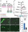
FIGURE 2:
Double-null animals show defects in cell proliferation and gene expression in the postembryonic mesoderm. (A–B) The C. elegans hermaphrodite postembryonic M lineage (modified from Jiang et al., 2005). Times indicated are hours posthatching at 25°C. (A) The M lineage with all the differentiated cell types; corresponding stages referred to in the text are indicated at left. (B) A schematic lateral view of the M lineage through larval development. D, dorsal; V, ventral; L, left; R, right; A, anterior; P, posterior. (C and D) Twenty-four hours after plating synchronized L1s, hypomorphic animals (C, with bright pharyngeal GFP) reached the 14-M stage, whereas double-null animals (D) were still at the 2-M stage. M lineage cells are marked by hlh-8::gfp (*). White arrowheads point to embryonically derived CCs. (E and F) hlh-8::gfp expression level at the 2-M stage is higher in a hypomorphic animal (E) than in a double-null animal (F). (G) Quantification of hlh-8::gfp signals at the 2-M stage. For each genotype, the pixel intensities of GFP signals from two M lineage cells in 10 different animals (20 cells total per genotype) were measured using Openlab software. Error bars represent 95% confidence intervals for the GFP intensity. Statistical significance was analyzed by Student's t test. ***, p < 0.001. Scale bars: 50 μm.
FIGURE 3:
FIGURE 3:
Nuclear and chromatin structure defects in dividing double-null cells. Indirect immunofluorescence images of the gonads (L2), hypodermis (L2), head (L2), and muscle cells (day 3) of animals grown at 20°C and stained with antibodies specific for Ce-lamin (Lamin) or FG-repeat–containing NPC proteins (414). Chromatin was stained with 4′,6-diamidino-2-phenylindole (DAPI). Arrows indicate abnormally condensed chromatin. Merged images shown at right; arrowheads indicate region shown at higher magnification (inset). Scale bars: 10 μm (applies to all panels).
FIGURE 4:
FIGURE 4:
Effects of LEM-domain mutations on C. elegans motility. Motility was measured as the number of head movements per minute on agar plates as a function of age (days 1, 2, 3, 4, 5, 6, 7, and 9 of development at 20°C), for wild-type (wt) animals, each indicated mutant strain, and double-null animals that expressed wild-type emr-1::GFP. Error bars indicate SEM. Significance was assessed by Student's t test (*, p < 0.05; **, p < 0.005; ***, p < 0.0005).
FIGURE 5:
FIGURE 5:
Thin-section TEM micrographs reveal abnormal muscle organization in double-null animals. Images of longitudinal sections from the midbody region at day 3 (A, D, and J) or day 6 (B and E), and transverse sections from a midbody region at day 6 (C, F, and K) of development at 20°C. Muscle organization appeared normal on days 3 and 6 in wild-type (WT) (A,B,C) and double-null animals expressing Ce-emerin::GFP on day 6 (J and K) but was abnormal in double-null animals (arrows in D and E). Also shown are longitudinal head sections (G and H) and transverse tail sections (I) from day 6 (G) or day 9 (H and I), in which sarcomeres were sometimes misoriented (*). Scale bars: 200 nm (A, D, C, and F), 500 nm (B, E, G, H, J, and K) or 5 µm (I).
FIGURE 6:
FIGURE 6:
Abnormal muscle cell and FO organization in double-null animals. (A) MYO-3::GFP expression in wild-type (wt) L2 stage animals (day 1), and double-null (emr-1−/−; lem-2−/−) animals at day 6 of development. Double-null muscle cells from the midbody to the tail were shorter (arrowheads). Scale bar: 10 μm. (B) Quantification of absolute muscle cell length (top), or after muscle cell length was normalized to animal length (bottom). (C) IFB-1::GFP expression in wild-type (wt) L2 stage animals on day 1 and double-null (emr-1−/−; lem-2−/−) animals on day 3 of development. The figure shows the posterior half of the animal. Double-null animals showed abnormal distribution of IFB-1::GFP in all the hypodermal cells in this region. Scale bar: 10 μm.
FIGURE 7:
FIGURE 7:
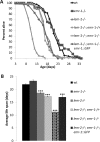
Shorter life span in animals lacking Ce-emerin and LEM-2. (A) Survival plots of wild-type (N2) animals (wt), and animals with each mutant genotype, at 20°C (see Materials and Methods). (B) Data from (A) were used to calculate average life span. Exogenous Ce-emerin::GFP expression restored the average life span of double-null animals to that of animals with one copy of emr-1 (emr-1−/+; lem-2−/−). (In a comparison with wt, *** represents p < 0.0001.)
FIGURE 8:
FIGURE 8:
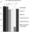
Effects of LEM-domain mutations on C. elegans smooth muscle activity. Pharynx pumping activity (pumps/min) was counted for 1 min in animals (n > 30 for each strain) at day 3 of development at 20°C when all animals were young adults, except the double-nulls, which arrested at L2. Expression of Ce-emerin::GFP rescued the pumping defect in double-null animals. Error bars indicate SEM. Significance was assessed by Student's t test (*, p < 0.05; **, p < 0.005; ***, p < 0.0005).

References

    1. Bione S, Maestrini E, Rivella S, Mancini M, Regis S, Romeo G, Toniolo D. Identification of a novel X-linked gene responsible for Emery-Dreifuss muscular dystrophy. Nat Genet. 1994;8:323–327. - PubMed
    1. Brenner S. The genetics of Caenorhabditis elegans. Genetics. 1974;77:71–94. - PMC - PubMed
    1. Chisholm AD, Hardin J. Epidermal morphogenesis. In: Martin Chalfie., editor. WormBook: The Online Review of C. elegans Biology. 2005. DOI: 10.1895/wormbook.1.35.1. - PMC - PubMed
    1. Cohen M, Tzur YB, Neufeld E, Feinstein N, Delannoy MR, Wilson KL, Gruenbaum Y. Transmission electron microscope studies of the nuclear envelope in Caenorhabditis elegans embryos. J Struct Biol. 2002;140:232–240. - PubMed
    1. Corsi AK, Kostas SA, Fire A, Krause M. Caenorhabditis elegans Twist plays an essential role in non-striated muscle development. Development. 2000;127:2041–2051. - PubMed

Publication types

MeSH terms

LinkOut - more resources