. 2011 Dec;134(Pt 12):3590-601.
doi: 10.1093/brain/awr307.

Quantification of increased cellularity during inflammatory demyelination

Affiliations

Quantification of increased cellularity during inflammatory demyelination

Yong Wang et al. Brain. 2011 Dec.

Abstract

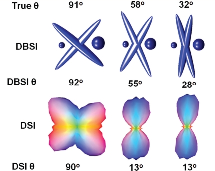
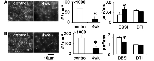
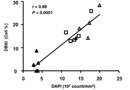
Multiple sclerosis is characterized by inflammatory demyelination and irreversible axonal injury leading to permanent neurological disabilities. Diffusion tensor imaging demonstrates an improved capability over standard magnetic resonance imaging to differentiate axon from myelin pathologies. However, the increased cellularity and vasogenic oedema associated with inflammation cannot be detected or separated from axon/myelin injury by diffusion tensor imaging, limiting its clinical applications. A novel diffusion basis spectrum imaging, capable of characterizing water diffusion properties associated with axon/myelin injury and inflammation, was developed to quantitatively reveal white matter pathologies in central nervous system disorders. Tissue phantoms made of normal fixed mouse trigeminal nerves juxtaposed with and without gel were employed to demonstrate the feasibility of diffusion basis spectrum imaging to quantify baseline cellularity in the absence and presence of vasogenic oedema. Following the phantom studies, in vivo diffusion basis spectrum imaging and diffusion tensor imaging with immunohistochemistry validation were performed on the corpus callosum of cuprizone treated mice. Results demonstrate that in vivo diffusion basis spectrum imaging can effectively separate the confounding effects of increased cellularity and/or grey matter contamination, allowing successful detection of immunohistochemistry confirmed axonal injury and/or demyelination in middle and rostral corpus callosum that were missed by diffusion tensor imaging. In addition, diffusion basis spectrum imaging-derived cellularity strongly correlated with numbers of cell nuclei determined using immunohistochemistry. Our findings suggest that diffusion basis spectrum imaging has great potential to provide non-invasive biomarkers for neuroinflammation, axonal injury and demyelination coexisting in multiple sclerosis.


Figures

Figure 1
Figure 1
Coronal images of the mouse brain were obtained from the sagittal scout (A). The width of the yellow rectangles represents the actual slice thickness of the image. The immunohistochemistry was performed on the sagittal slices for the ease of axon and myelin counting. The corresponding immunohistochemistry counting regions (white rectangles) are displayed with the location and thickness of one image voxel (yellow squares; B). In vivo fractional anisotropy maps of rostral (C), middle (D) and caudal (E) corpus callosum (from a control mouse; colour-coded full view) reveal the structure of white matter tracts. The region of interest for DBSI analysis is marked in yellow rectangles shown in the expanded view for both control and cuprizone-fed mice.
Figure 2
Figure 2
The representative result of DAPI (A) and SMI-31 (B) staining from a fixed mouse trigeminal nerve. Both nucleus and axon counts were performed using CellC program (C). The fraction of MRI-derived cell and fibre component was determined by DBSI (D). The ratio of nucleus and axon counts by immunohistochemistry (IHC) (6.20 ± 1.81) was comparable to the ratio of DBSI-derived cell and fibre percentage (6.26 ± 0.93; n = 8). DBSI = diffusion basis spectrum imaging.
Figure 3
Figure 3
DTI failed to correctly estimate the diffusion parameters (λ||, open circles; λ, open squares; fractional anisotropy, open triangles) due to the partial volume effect of the added gel (the more gel added the more significant deviation). DBSI correctly estimated diffusion parameters (λ||, solid circles; λ, solid squares; fractional anisotropy, solid triangles) by separating and quantifying the gel (A). Comparable λ|| (B) and λ (C) derived from trigeminal nerves with and without gel were further confirmed by Bland–Altman analysis. Representative DBSI-derived isotropic diffusion spectra for fibre only (grey dashed line) and for fibre with gel phantom (black solid line) are compared (D). The diffusivity component near 0 µm2/ms was assigned to the cell. The higher diffusivity component in fibre only (grey dashed line) was assigned to the inter-axonal and extracellular water. The higher diffusivity component in the fibre with gel (black solid line) was assigned to inter-axonal, extracellular and free diffusion gel water. DBSI-derived gel fractions agreed with that determined by T2-weighted intensity fractions (D inset). DTI = diffusion tensor imaging; DBSI = diffusion basis spectrum imaging; FA = fractional anisotropy.
Figure 4
Figure 4
Resolution of crossing fibres by diffusion spectrum imaging and DBSI. Although, the goal of this study was to demonstrate the confounding effect of increased cellularity in white matter lesions, we would also like to demonstrate the capability of DBSI to resolve crossing fibres deriving crossing angles, fraction of fibres and diffusion parameters of individual fibres. Gels were added to mimic oedema/CSF contamination/tissue loss. For 91°, 58° and 32° phantoms, DBSI determined angles were 92°, 55° and 28°; diffusion spectrum imaging-derived angles were 90°, 13° and 13°. DBSI also correctly recovered the directional diffusivity of the crossing fibres removing the effect of gel (λ|| = 1.14 ± 0.06 µm2/ms, and λ = 0.12 ± 0.02 µm2/ms). The fraction of the extent of gel was also estimated by DBSI.
Figure 5
Figure 5
The middle corpus callosum after 4 weeks of cuprizone treatment was examined using in vivo diffusion MRI (analysed using both DTI and DBSI model) followed by post-mortem immunohistochemistry using (A) the antibody against myelin basic protein, and (B) the antibody against phosphorylated neurofilament (SMI-31), to quantify the extent of demyelination (A), and axonal injury (B). All data were derived from the same region of interest from the middle corpus callosum as marked in Fig. 1B and D. Due to the partial volume effect originating from the surrounding grey matter, DTI failed to depict the extent of demyelination or axonal injury in this region, overestimating λ while underestimating λ|| of the control corpus callosum. The myelinated axon counts significantly decreased by 77% (65 609 ± 14 804 versus 14 830 ± 12 407 per mm2) in the cuprizone-treated mice compared with that of the control. The λ increased by 70% measured using DBSI (0.31 ± 0.05 versus 0.51 ± 0.09 µm2/ms) in the cuprizone-treated mice compared with that of the control. However, DTI determined λ values were not different between the cuprizone-treated (0.43 ± 0.03 µm2/ms) and the control mice (0.47 ± 0.04 µm2/ms). The SMI-31-positive axon counts significantly decreased in the cuprizone-treated corpus callosum (155 832 ± 29 934 versus 53 685 ± 18 712 per mm2) compared with that of the control. The DBSI derived λ|| significantly decreased by 28% (2.05 ± 0.17 versus 1.47 ± 0.09 µm2/ms) while no difference was observed in that derived using DTI (1.14 ± 0.09 versus 1.11 ± 0.03 µm2/ms) in the cuprizone-treated mice compared with that of the control.
Figure 6
Figure 6
Cell densities quantified based on DAPI-positive nucleus counts linearly correlating with the restricted diffusion (assigned to cells) ratio derived by DBSI. Data from both the control group (caudal corpus callosum: filled triangle) and the 4-week cuprizone-treated group (caudal corpus callosum: open triangle; middle corpus callosum: open square) were analysed by linear regression. The significant correlation (r = 0.86, P < 0.0001) supported that DBSI-derived cell ratio can potentially be used as a novel non-invasive index of inflammation or gliosis. DBSI = diffusion basis spectrum imaging.
Figure 7
Figure 7
Different white matter pathologies associated multiple tensor representation and DTI simplification. Grey ellipsoid represents the diffusion tensor profile for normal myelinated white matter with λ|| > λ. Black drawings represent the diffusion profiles for multiple tensor representation or DTI simplification: (A) co-existing axon and myelin injury of coherent pure myelinated axons; (B) axon and myelin injury with cell infiltration; (C) axon and myelin injury with axonal loss; and (D) axon and myelin injury, cell infiltration or proliferation and axonal loss.

References

    1. Alexander DC. Multiple-fiber reconstruction algorithms for diffusion MRI. Ann N Y Acad Sci. 2005;1064:113–33. - PubMed
    1. Alexander DC, Hubbard PL, Hall MG, Moore EA, Ptito M, Parker GJ, et al. Orientationally invariant indices of axon diameter and density from diffusion MRI. Neuroimage. 2010;52:1374–89. - PubMed
    1. Anderson AW. Measurement of fiber orientation distributions using high angular resolution diffusion imaging. Magn Reson Med. 2005;54:1194–206. - PubMed
    1. Anderson AW, Xie J, Pizzonia J, Bronen RA, Spencer DD, Gore JC. Effects of cell volume fraction changes on apparent diffusion in human cells. Magn Reson Imaging. 2000;18:689–95. - PubMed
    1. Audet C, Dennis JE. Analysis of generalized pattern searches. Siam J Optimization. 2003;13:889–903.

Publication types