

doi: 10.1186/1746-4811-7-46.
Reproducible hairy root transformation and spot-inoculation methods to study root symbioses of pea
Affiliations
- PMID: 22172023
- PMCID: PMC3264533
- DOI: 10.1186/1746-4811-7-46
Item in Clipboard
Reproducible hairy root transformation and spot-inoculation methods to study root symbioses of pea
Plant Methods.
.
Abstract
Pea has lagged behind other model legumes in the molecular study of nodulation and mycorrhizae-formation because of the difficulty to transform its roots and its poor growth on agar plates. Here we describe for pea 1) a transformation technique which permits the complementation of two known non-nodulating pea mutants, 2) a rhizobial inoculation method which allows the study of early cellular events giving rise to nodule primordia, and 3) a targeted fungal inoculation method which allows us to study short segments of mycorrhizal roots assured to be infected. These tools are certain to advance our knowledge of pea root symbioses.
Figures
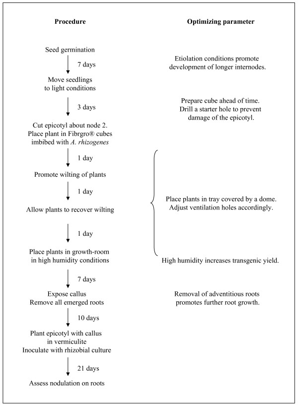
Schematic representation of the time-line required for the generation of composite pea plants, with an emphasis on the parameters which must be optimized.
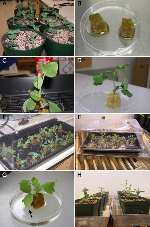
Illustrations depicting the main steps for the production of composite pea plants. A. Plants grown for 10 days in vermiculite (7 days in dark followed by 3 in light). B. Fibrgro® cubes saturated with A. rhizogenes. C. A shoot of a 10 day-old plant cut above node 2 and inserted into Fibrgro® cube. D. A wilted pea plant about to receive water for recovery. E. Plants placed in growth tray under a plastic dome. F. High humidity is provided by plastic lid with closed ventilation holes and metal tray with water. G. A root (arrow) protruding through a Fibrgro® cube 10 days after A. rhizogenes infection. H. Callus-forming plants, the regenerated roots of which have been excised, placed in an upright position in vermiculite before being inoculated with R. leguminosarum 3 days later.
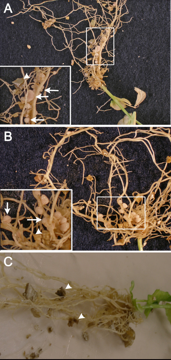
Macro-photography of developing nodules on transformed roots. Non-nodulation mutants P56 (A) and P5 (B) had their nodulation capabilities restored through A. rhizogenes transformation. Each inset shows a magnification of the area framed in the main photograph. Root nodules (arrows) that have emerged from the transformed roots should not be confused with vermiculite pieces (arrowheads). C. P56 mutant transformed with AR1193 lacking the sym10 construct did not exhibit any nodules.
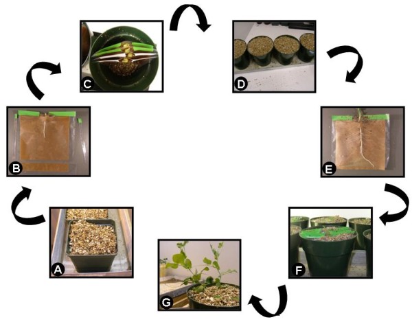
Schematic representation of the important steps for spot-inoculation. A. Imbibed seeds placed in vermiculite. B. Seedling radicle placed in trough of trimmed pouch. C. Pea cotyledons and epicotyls protruding from tightly-closed pouches. D. Pots, with pouches inside, filled with vermiculite to prevent light exposure. E. Inoculation 5 days later with R. leguminosarum bv. vicieae placed as a minute drop (seen as a black dot drawn with a marker on plastic) at the most susceptible zone of infection of several lateral roots. F. Pouches later returned to pots. G. Healthy plants allowed to grow until inspection.
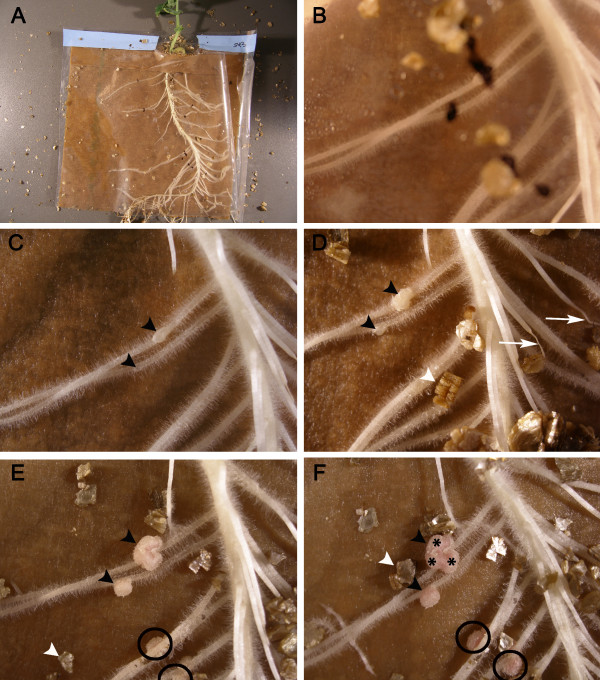
Macro-photography of nodules developing on roots of plants grown in pouches. A. An overview of an 8 day-old spot-inoculated plant. B. A close-up focussed on the root hairs of the seedling 5 DAI. The unfocussed black dots are located on the surface of the pouch and correspond to the different inoculation sites. In C to F, two nodules (arrowheads) can be seen developing over time. The nodules are five-day old (C), seven-day old (D), 10 day-old (E) and 14 day-old (F). In E and F, two additional nodules (black circles) appeared later at the spot-inoculation site; they cannot be given a specific age. One (asterisks in F) of the nodules appears much larger than the others; it is evident that this large nodule has multiple meristems. In D, two roots (arrows) which did not grow in contact with the filter paper of the pouch dried out. White arrowheads indicate pieces of vermiculite.
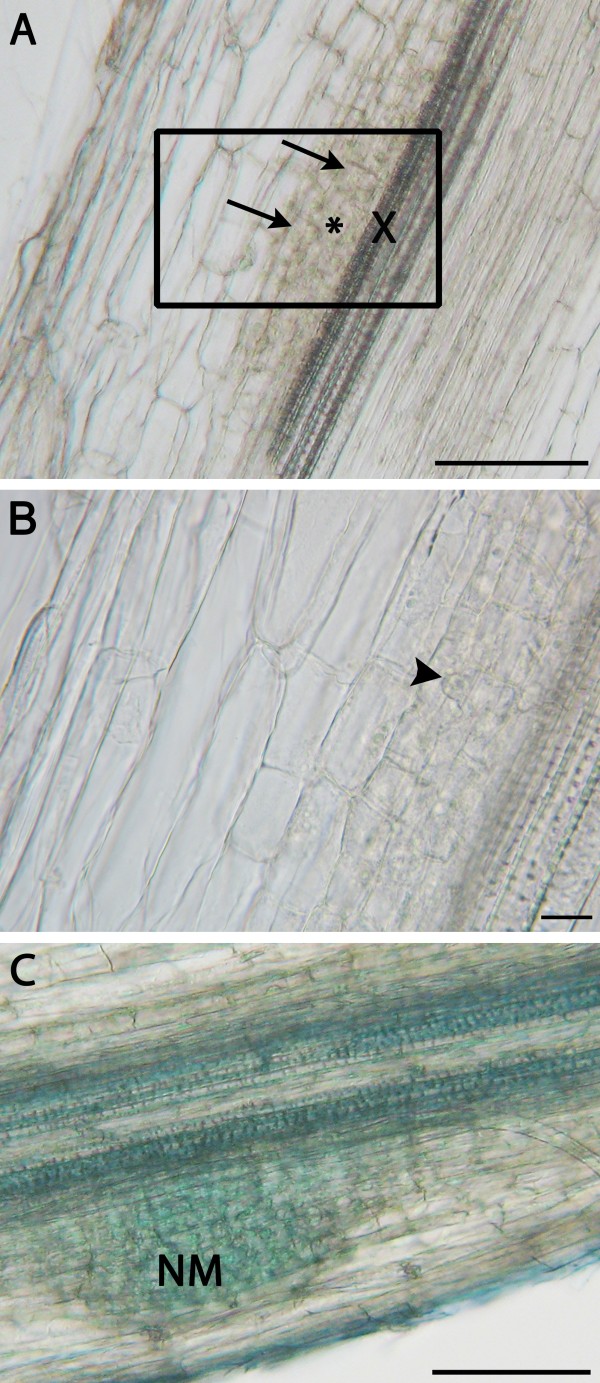
Microscopy of spot-inoculated lateral roots. A. An unstained longitudinal section of a spot-inoculated lateral root 24 hours after rhizobial inoculation showing anticlinal divisions in its inner cortex (arrows), and in its pericycle (asterisk), located adjacent to a xylem pole (X). Bar: 100 μm. B. A close-up on the area framed in Figure A. Metabolically active cells, one with a large nucleus (arrowhead), exhibit anticlinal divisions. Bar: 10 μm. C. A longitudinal section of a root inoculated 3 days earlier with rhizobia and stained with toluidine blue. The nodule meristem (NM) has passed the mid-point of the root cortical region. Bar: 100 μm. Note that for all the photographs, there was no indication of nodule emergence and it was only because of the spot-inoculation that these events were easily captured. All sections were made with a vibratome.
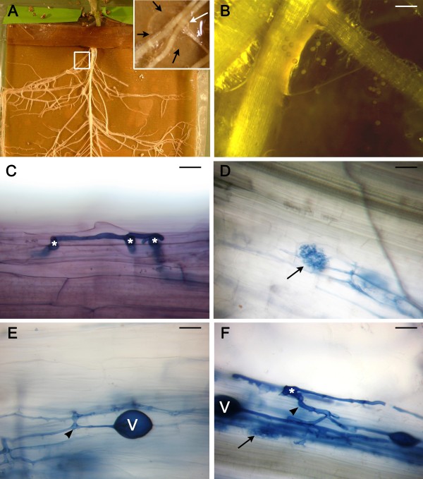
Fungal spot-inoculation of plants grown in a pouch. A. An overview of the root system of a 40-day old plant grown in a pouch. The plant was challenged when it was 5 day-old with fungal spores located in an agar plug. Inset: A close-up of the area indicated by a white square in A. The plug (delineated by arrows) was placed below the lateral roots. B. Close up on the roots which were pushed gently into the plug containing the fungal spores. Bar: 0.5 mm. C. An extra-radical hypha and some hyphopodia (asterisks) on the surface of a cleared root inoculated 21 days earlier. D. Arbuscules located within inner cortical cells of a cleared root inoculated 35 days earlier. E. Intraradical hyphae and a vesicle in the cortex of a root 35 DAI. F. An infection unit with extensive hyphal branches; note the hyphopodium (asterisk), the main intraradical hypha (arrowhead), an arbuscule (arrow), and a vesicle (V). C - F: Bar: 20 μm; ink-vinegar staining.
Similar articles
-
Agrobacterium rhizogenes-mediated transformation of Pisum sativum L. roots as a tool for studying the mycorrhizal and root nodule symbioses.PeerJ. 2019 Mar 6;7:e6552. doi: 10.7717/peerj.6552. eCollection 2019. PeerJ. 2019. PMID: 30863680 Free PMC article.
-
Common and divergent shoot-root signalling in legume symbioses.New Phytol. 2016 Apr;210(2):643-56. doi: 10.1111/nph.13779. Epub 2015 Dec 11. New Phytol. 2016. PMID: 26661110
-
The application of CRISPR/Cas9 in hairy roots to explore the functions of AhNFR1 and AhNFR5 genes during peanut nodulation.BMC Plant Biol. 2020 Sep 7;20(1):417. doi: 10.1186/s12870-020-02614-x. BMC Plant Biol. 2020. PMID: 32894045 Free PMC article.
-
[Comparative genetics and evolutionary morphology of symbiosis formed by plants with nitrogen-fixing microbes and endomycorrhizal fungi].Zh Obshch Biol. 2002 Nov-Dec;63(6):451-72. Zh Obshch Biol. 2002. PMID: 12510586 Review. Russian.
-
The Role of Gibberellins and Brassinosteroids in Nodulation and Arbuscular Mycorrhizal Associations.Front Plant Sci. 2019 Mar 15;10:269. doi: 10.3389/fpls.2019.00269. eCollection 2019. Front Plant Sci. 2019. PMID: 30930916 Free PMC article. Review.
Cited by
-
Use of ex vitro composite plants to study the interaction of cowpea (Vigna unguiculata L.) with the root parasitic angiosperm Striga gesnerioides.Plant Methods. 2012 Jun 28;8(1):22. doi: 10.1186/1746-4811-8-22. Plant Methods. 2012. PMID: 22741546 Free PMC article.
-
Dryas as a Model for Studying the Root Symbioses of the Rosaceae.Front Plant Sci. 2019 Jun 4;10:661. doi: 10.3389/fpls.2019.00661. eCollection 2019. Front Plant Sci. 2019. PMID: 31214211 Free PMC article.
-
Genotype-independent Agrobacterium rhizogenes-mediated root transformation of chickpea: a rapid and efficient method for reverse genetics studies.Plant Methods. 2018 Jul 6;14:55. doi: 10.1186/s13007-018-0315-6. eCollection 2018. Plant Methods. 2018. PMID: 29988950 Free PMC article.
-
Gene-based SNP discovery and genetic mapping in pea.Theor Appl Genet. 2014 Oct;127(10):2225-41. doi: 10.1007/s00122-014-2375-y. Epub 2014 Aug 15. Theor Appl Genet. 2014. PMID: 25119872 Free PMC article.
-
An anthocyanin marker for direct visualization of plant transformation and its use to study nitrogen-fixing nodule development.J Plant Res. 2019 Sep;132(5):695-703. doi: 10.1007/s10265-019-01126-6. Epub 2019 Jul 19. J Plant Res. 2019. PMID: 31325057 Free PMC article.
References
-
- Ellis THN, Poyser SJ. An integrated and comparative view of pea genetic and cytogenetic maps. New Phytol. 2002;153:17–25. doi: 10.1046/j.0028-646X.2001.00302.x. - DOI
-
- Dolgikh EA, Leppyanen IV, Osipova MA, Savelyeva NV, Borisov AY, Tsyganov VE, Geurts R, Tikhonovich IA. Genetic dissection of Rhizobium-induced infection and nodule organogenesis in pea based on ENOD12A and ENOD5 expression analysis. Plant Biol. 2011;13:285–296. doi: 10.1111/j.1438-8677.2010.00372.x. - DOI - PubMed
-
- Szczyglowski K, Stougaard J. Lotus genome: pod of gold for legume research. TIPS. 2008;13:515–517. - PubMed
-
- Timmers ACJ, Auriac M-C, Truchet G. Refined analysis of early symbiotic steps of the Rhizobium-Medicago interaction in relationship with microtubular cytoskeleton rearrangements. Development. 1999;126:3617–3628. - PubMed
LinkOut - more resources
Full Text Sources
