. 2011 Nov-Dec;58(6):391-6.
doi: 10.1002/bab.52. Epub 2011 Oct 28.

Biochemical characterization of fluorescent-labeled recombinant human alpha-L-iduronidase in vitro

Affiliations

Biochemical characterization of fluorescent-labeled recombinant human alpha-L-iduronidase in vitro

Brigette L Tippin et al. Biotechnol Appl Biochem. 2011 Nov-Dec.

Abstract

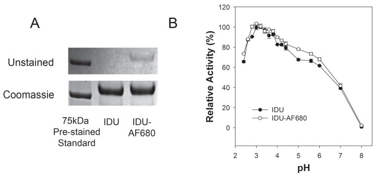
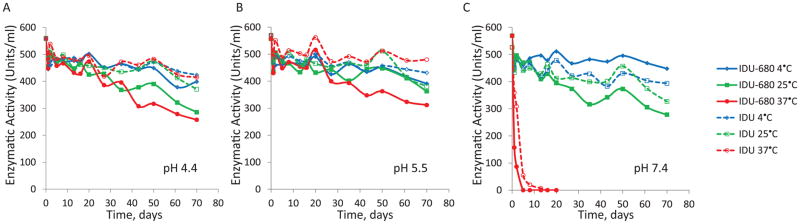
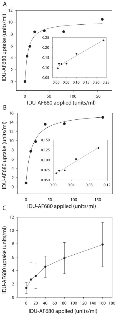
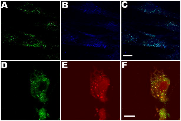
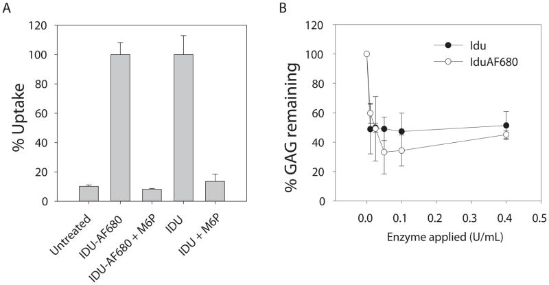
In vivo tracking of the delivery of therapeutic proteins is a useful tool for preclinical studies. However, many labels are too large to use without disrupting the normal uptake, function, or other properties of the protein. Low-molecular-weight fluorescent labels allow in vivo and ex vivo tracking of the distribution of therapeutic proteins, and should not alter the protein's characteristics. We tested the in vitro properties of fluorescent-labeled recombinant human alpha-l-iduronidase (rhIDU, the enzyme deficient in Hurler syndrome) and compared labeled to unlabeled proteins. Labeled rhIDU retained full enzymatic activity and showed similar kinetics to nonlabeled rhIDU. Uptake of labeled rhIDU into human Hurler fibroblasts, measured by activity assay, was equivalent to unlabeled rhIDU enzyme and showed an uptake constant of 0.72 nM. Labeled rhIDU was also able to enter cells via the mannose 6-phospate receptor pathway and reduce glycosaminoglycan storage in Hurler fibroblasts. Subcellular localization was verified within lysosomes by confocal microscopy. These findings suggest that fluorescent labeling does not significantly interfere with enzymatic activity, stability, or uptake, and validates this method as a way to track exogenously administered enzyme.


Conflict of interest statement

Conflicts of interest: The Los Angeles Biomedical Research Institute at Harbor-UCLA Medical Center has a financial interest in recombinant human alpha-L-iduronidase (laronidase). Dr. Dickson receives research support from Biomarin Pharmaceuticals, Inc. and Genzyme Corporation.

Figures

Figure 1
Figure 1
Labeling and pH optimum of rhIDU. (A) Polyacrylamide gel electrophoresis (4–20%) analysis of rhIDU (IDU) before and after the labeling process. Upper panel shows unstained gel with Alexa Fluor 680 labeled protein (IDU-AF680) clearly visible to the naked eye (DOL of 3.1). Lower panel is the same gel after staining with coomassie blue. (B) Labeled (DOL 3.1) or unlabeled IDU were diluted to 16 μg/ml in 200 mM/100 mM phosphate-citrate buffer at the indicated pHs (2.4–8.0) and immediately assayed for enzyme activity towards the artificial fluorogenic substrate 4-MUI for 15 minutes at 22 °C. Relative activity for each enzyme was plotting from an average of three independent experiments where the maximally observed activity was defined as 100%.
Figure 2
Figure 2
Stability of fluorescent-labeled rhIDU. Stability of the enzyme as a function of pH and storage temperature for labeled and unlabeled rhIDU are shown over the course of 70 days incubation. Labeled (solid lines) and unlabeled (dashed lines) enzymes were diluted to 16 μg/ml in 100 mM/50 mM phosphate-citrate buffer at pH 4.4 (A), pH 5.5 (B), and pH 7.4 (C) and these stock solutions were incubated at various temperatures, 4°C (blue), 25°C (green), 37°C (red) over a 70-day period. Enzyme aliquots were taken throughout this period of time and activity was estimated under the standard conditions (25 mM 4-MUI for 10 minutes at 22 °C). Units of activity are defined as nmoles of 4-MU product formed per hour.
Figure 3
Figure 3
Kinetics of cellular uptake of rhIDU labeled with Alexa Fluor 680. Labeled rhIDU (IDU-AF680) at either DOL of 1.7 (A) or DOL 3.1 (B) was applied to confluent Hurler fibroblasts (GM 1391) in 6 well dishes at the indicated doses in MEM medium without serum and incubated for 4 hours. Double reciprocal (Lineweaver-Burk) plots of the kinetic data are shown as insets. (C) Qualitative direct detection of Alexa Fluor 680 taken up by Hurler cells. Fluorescence was monitored in cell lysates at 640/700 nm excitation/emission wavelengths, and uptake was determined by comparison to a standard curve of fluorescent intensity emitted at 700 nm vs. input amount of Idu-AF680 (units/ml). All experiments were performed in triplicate.
Figure 4
Figure 4
Uptake and lysosomal targeting of fluorescent-labeled rhIDU into human Hurler patient fibroblast cultures. AF680 labeled enzyme (160 units) was directly applied to Hurler fibroblasts grown on coverslips in serum-free MEM and incubated for 4 hours at 37 °C and 5% CO2. (A–C) Detection of AF680 label (blue) anti-rhIDU antibodies (green) and co-localization (aqua) in Hurler cells. (D–F) Lysosomal targeting of rhIDU-AF680. Detection with anti-iduronidase antibody (green), LysoTracker Red DND-99 (red), and merge. All colors are generated by Leica LCS imaging software. Scale bars are 15 μm.
Figure 5
Figure 5
Mannose 6-phosphate receptor mediated delivery and GAG storage reduction. (A) 80 activity units per ml rhIDU with Alexa Fluor 680 label (IDU-AF680) or unlabeled (IDU) were applied to confluent Hurler fibroblasts in six-well plates containing 1 ml of serum free MEM with or without competing 5 mM mannose 6-phosphate (M6P) for 4 hours. Enzymatic activity assays using 4-MUI substrate were performed on cell lysates and percent uptake was plotted relative to maximal uptake observed in the absence of inhibitor. (B) IDU or IDU-AF680 was applied (0.0–0.4 units/ml) to cultured Hurler fibroblasts in the presence of H235SO4 and incubated for 48 h at 37 °C and 5% CO2 as described in the methods and reported in detail previously [11]. Radioactive labeled GAG were isolated from harvested cell pellets by two rounds of ethanol extraction, acid solubilized, and quantified by scintillation counting. Radioactive counts per minute were normalized to total protein concentration in the extracted pellet and plotted as relative percent labeled GAG compared to the untreated samples at 0 units of enzyme applied.

References

    1. Leader B, Baca QJ, Golan DE. Nat Rev Drug Discovery. 2008;7:21–39. - PubMed
    1. Barton NW, Brady RO, Dambrosia JM, Di Bisceglie AM, Doppelt SH, Hill SC, Mankin HJ, Murray GJ, Parker RI, Argoff CE, Grewal RP, Yu KT. N Engl J Med. 1991;324:1464–1470. - PubMed
    1. Eng CM, Guffon N, Wilcox WR, Germain DP, Lee P, Waldek S, Caplan L, Linthorst GE, Desnick RJ the International Fabry Disease Study Group. N Engl J Med. 2001;345:9–16. - PubMed
    1. Kakkis ED, Muenzer J, Tiller GE, Waber L, Belmont J, Passage M, Izykowski B, Phillips J, Doroshow R, Walot I, Hoft R, Neufeld E. N Engl J Med. 2001;344:182–188. - PubMed
    1. Harmatz P, Giugliani R, Schwartz I, Guffon N, Teles EL, Miranda MCS, Wraith JE, Beck M, Arash L, Scarpa M, Yu ZF, Wittes J, Berger KI, Newman MS, Lowe AM, Kakkis E, Swiedler SJ. J Pediatrics. 2006;148:533–539. - PubMed

Publication types

LinkOut - more resources