. 2012 Jan;11(1):87-97.
doi: 10.1158/1535-7163.MCT-11-0540. Epub 2011 Dec 15.

SOD mimetics: a novel class of androgen receptor inhibitors that suppresses castration-resistant growth of prostate cancer

Affiliations

SOD mimetics: a novel class of androgen receptor inhibitors that suppresses castration-resistant growth of prostate cancer

Rusha Thomas et al. Mol Cancer Ther. 2012 Jan.

Abstract

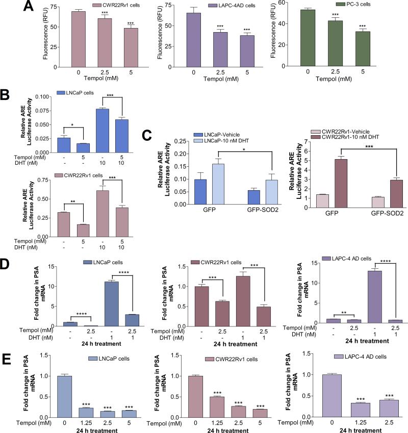
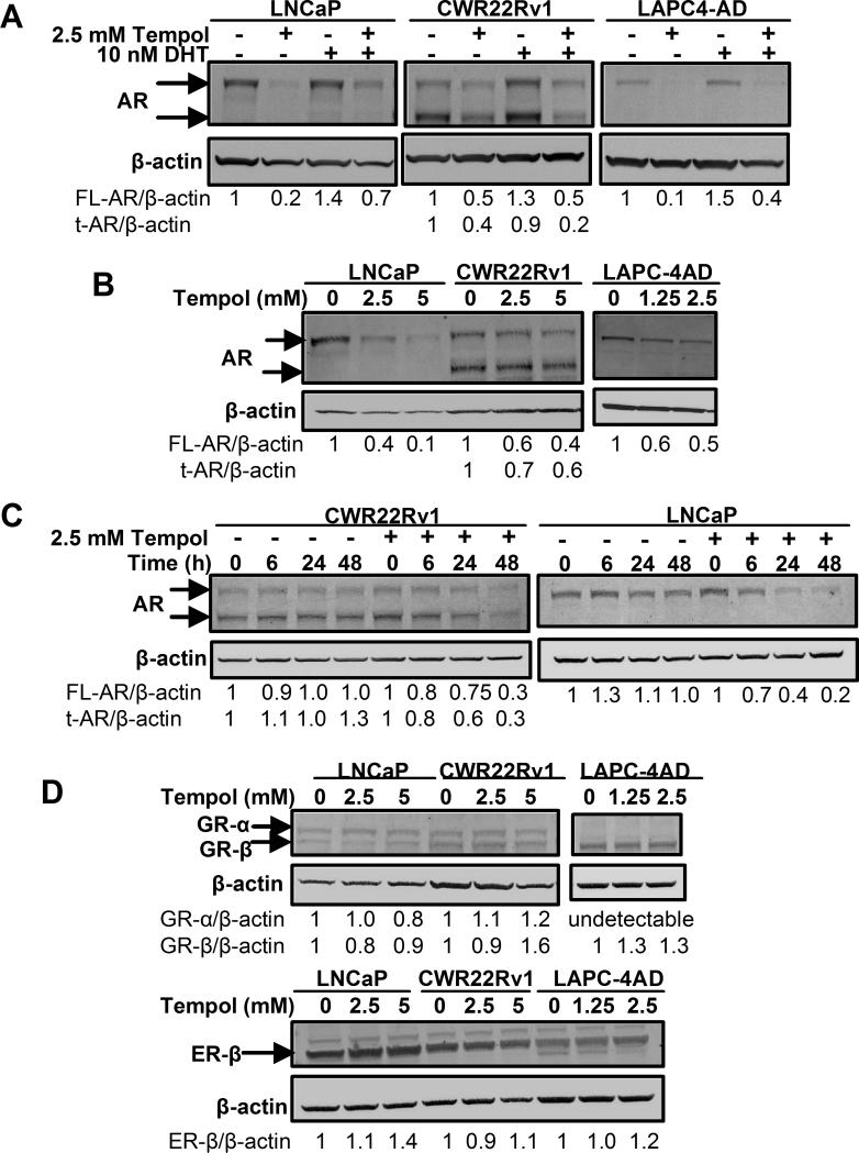
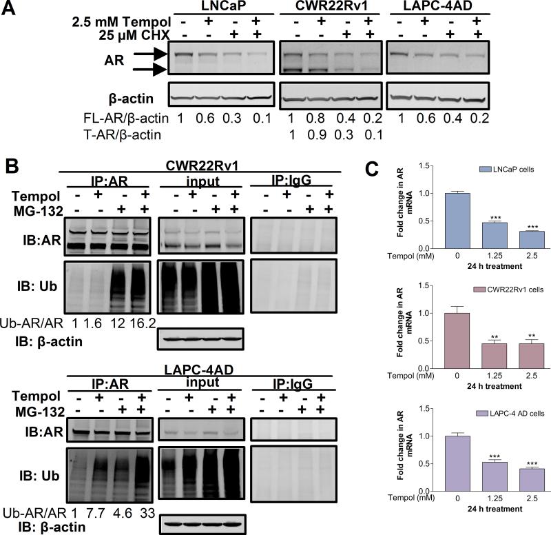
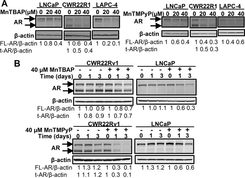
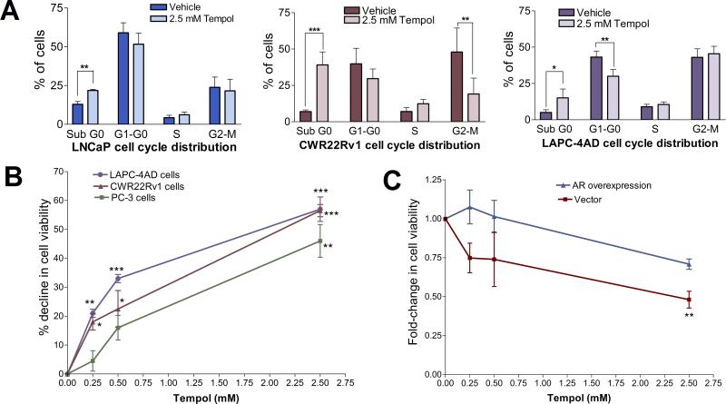
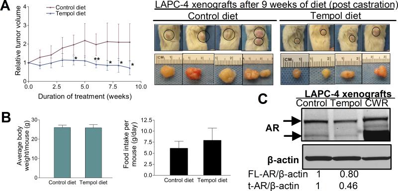
Advanced prostate cancer is the second leading cause of cancer-related deaths among American men. The androgen receptor (AR) is vital for prostate cancer progression, even in the face of castrate levels of serum testosterone following androgen ablation therapy, a mainstay therapy for advanced prostate cancer. Downregulation of superoxide dismutase 2 (SOD2), a major intracellular antioxidant enzyme, occurs progressively during prostate cancer progression to advanced states and is known to promote AR activity in prostate cancer. Therefore, this study investigated the effects of SOD mimetics on AR expression and function in AR-dependent LNCaP, CWR22Rv1, and LAPC-4AD prostate cancer cells. Treatment with Tempol (4-hydroxy-2,2,6,6-tetramethylpiperidine-N-oxyl), a SOD mimetic, not only lowered cellular superoxide levels but also concomitantly attenuated AR transcriptional activity and AR target gene expression in a dose- and time-dependent manner, in the presence and absence of dihydrotestosterone, the major endogenous AR agonist. Inhibition of AR by Tempol was mediated, in large part, by its ability to decrease AR protein via increased degradation, in the absence of any inhibitory effects on other nuclear receptors. Inhibitory effects of Tempol on AR were also reproducible with other SOD mimetics, MnTBAP and MnTMPyP. Importantly, effects of Tempol on AR function were accompanied by significant in vitro and in vivo reduction in castration-resistant prostate cancer (CRPC) survival and growth. Collectively, this study has shown for the first time that SOD mimetics, by virtue of their ability to suppress AR function, may be beneficial in treating the currently incurable CRPC, in which SOD2 expression is highly suppressed.


Figures

Fig. 1
Fig. 1. Tempol-mediated decline in cellular O2- levels is accompanied by a significant reduction in AR transcriptional activity
(A) Tempol decreases cellular O2- levels. Cells were treated with 0-5 mM Tempol in phenol-red-free (PR-free), serum-free medium for 24 h, followed by O2- analysis with hydroethidine. Data are mean ± S.D. Statistical significance assessed by One-way ANOVA is versus untreated cells. (B) Tempol inhibits androgen response element (ARE)-driven luciferase reporter activity, both in the presence and absence of DHT. Cells were transiently co-transfected with plasmids pGL3-TK-3xARE-FLuc and pGL4.75-RLuc, followed by treatment with 5 mM Tempol +/- 10 nM DHT for 24 h. Relative luciferase activity was determined and represented as mean ± S.D. Statistical significance was assessed by Unpaired t test. (C) Tempol's effect on ARE-driven luciferase reporter activity is closely mimicked by SOD2 overexpression. Cells were transiently co-transfected with GFP-SOD2 or GFP plasmids, along with pGL3-TK-3xARE-FLuc and pGL4.75-RLuc constructs, and subsequently treated for 24 h with vehicle or 10 nM DHT. Relative luciferase activity was determined and represented as mean ± S.D. Statistical significance was assessed by Unpaired t test. (D) Tempol decreases PSA mRNA, both in the presence and absence of DHT. Cells were treated for 24 h with 1 nM DHT +/- 2.5 mM Tempol in PR-free medium containing 5% charcoal-stripped (C/S) FBS. PSA mRNA was assessed by qPCR and expressed as fold change +/- S.D. (ΔΔCt method), with mRNA levels in the vehicle-treated control set at 1. Statistical significance was assessed by Unpaired t test. (E) Tempol elicits a dose-dependent decline in PSA mRNA. Cells were treated for 24 h with increasing doses of Tempol in serum-free medium, followed by qPCR analyses of PSA mRNA, which was expressed as fold change +/- S.D., with mRNA levels in the untreated control set at 1. Statistical significance assessed by One-way ANOVA is versus the control. For Figs. 1A-1E, * indicates p<0.05, ** indicates p<0.01, *** indicates p<0.001, and **** indicates p<0.0001.
Fig.2
Fig.2. Tempol decreases AR protein, without significantly altering the expression of other nuclear receptors
(A) Tempol decreases AR protein, both in the presence and absence of DHT. Cells were grown in PR-free medium containing 5% C/S FBS for 2 d prior to 24-h treatment with 10 nM DHT +/- 2.5 mM Tempol. Cell lysates were prepared and AR protein analyzed by Western blotting. (B) Tempol elicits a dose-dependent decline in AR protein. Cells were treated for 24 h with increasing doses of Tempol in serum-free medium, followed by cell lysate preparation and AR protein analysis by Western blotting. (C) Tempol elicits a time-dependent decline in AR protein. Cells were treated with vehicle or 2.5 mM Tempol for 0-48 h in complete medium, followed by cell lysate preparation and AR protein analysis by Western blotting. AR expression was normalized to that of β-actin and expressed as fold-change, relative to that at the 0 h time point for Vehicle or 2.5 mM Tempol, respectively. (D) Tempol does not decrease protein levels of other nuclear receptors. Cells were treated for 24 h with increasing doses of Tempol in serum-free medium. Cell lysates were prepared and protein levels of nuclear receptors GR-α, GR-β and ER-β were assessed by Western blot analyses. For all Western blot analyses, nuclear receptor expression in each sample was normalized to that of the loading control, β-actin.
Fig.3
Fig.3. Tempol accelerates degradation of AR protein and reduces AR mRNA levels
(A) Tempol promotes AR protein degradation. Cells were treated with 25 μM CHX +/- 2.5 mM Tempol for 18 h in complete medium. CHX-treated samples were pre-treated with CHX for 6 h. AR expression was analyzed by Western blotting and expressed as fold-change, relative to vehicle control. (B) Tempol promotes ubiquitination of AR. Cells were treated with 5 mM Tempol +/- 2.5 μM MG-132 for 8 h, and AR was immunoprecipitated from the cell lysates. Samples immunoprecipitated with an isotype-matched control antibody were run in parallel. The immunoprecipitates and input lysates were subjected to Western blot analyses with antibodies specific for AR and ubiquitin (Ub). β-actin served as a loading control for the input. The intensity of ubiquitin in each immunoprecipitation sample was normalized to that of AR in the same sample and expressed as a fold-change, with the ubiquitinated AR/AR ratio in the untreated control set at 1. (C) Tempol dose-dependently decreases AR mRNA. Cells were treated for 24 h with increasing doses of Tempol in serum-free medium, followed by qPCR analyses of AR mRNA. AR mRNA levels were expressed as fold change +/- S.D. (ΔΔCt method), with mRNA levels in the untreated control set at 1. ** indicates p<0.01, and *** indicates p<0.001 versus the control (One-way ANOVA).
Fig.4
Fig.4. AR inhibition is also a property common to other SOD mimetics
(A) SOD mimetics, MnTBAP and MnTMPyP, elicit a dose-dependent decrease in AR protein. Cells were treated for 24 h with 0-40 μM MnTBAP or MnTMPyP in serum-free medium. AR protein was assessed by Western blotting. (B) MnTBAP and MnTMPyP decrease AR protein in a time-dependent manner. Cells were treated with vehicle or 40 μM MnTBAP or MnTMPyP, respectively, for 0-3 d in complete medium, and AR protein analyzed by Western blotting. The intensity of the AR protein band in each sample was normalized to that of β-actin in the same sample and expressed as a fold-change, relative to that at the 0 d time point for vehicle and 40 μM MnTBAP or MnTMPyP treatments, respectively.
Fig.5
Fig.5. Tempol decreases PCa cell viability
(A) Cell-cycle analysis of Tempol-treated cells reveals an increase in the dead cell fraction. Cells were treated with vehicle or 2.5 mM Tempol in PR-free medium containing 5% C/S FBS for 3 d, and cell-cycle distribution assessed by flow cytometry. Data are mean ± S.D., statistical significance was assessed by Unpaired t test. (B) Tempol elicits a dose-dependent decline in cell viability. Cells were treated with 0-2.5 mM Tempol in PR-free medium containing 5% C/S FBS for 2 d. Thereafter, cell viability was assessed and represented as % decline, relative to vehicle control. Data are mean ± S.D., and statistical significance was assessed by One-way ANOVA. (C) AR overexpression blunts Tempol-mediated decrease in PCa cell viability. LAPC-4AD cells were transfected with AR cDNA or empty vector, and treated for 1 d with 0-2.5 mM Tempol in PR-free medium containing 5% C/S FBS. Cell viability was assessed and expressed as fold-change relative to vehicle treatment. Data are mean ± S.D., and statistical significance was assessed by One-way ANOVA. For Figs. 5A-5C, * indicates p<0.05, ** indicates p<0.01, and *** indicates p<0.001 versus control.
Fig.6
Fig.6. Tempol decreases AR protein in vivo and suppresses growth of castration-resistant prostate cancer xenografts
(A, left panel) Tempol significantly suppresses growth of castration-resistant LAPC-4 PCa xenografts. Subcutaneous LAPC-4AD tumor-bearing mice were surgically castrated and placed on control diet (n=6) or Tempol diet (n=4) for 9 weeks. Mean relative tumor volume +/- S.D. for each group was calculated at the end of each week of treatment. * indicates p<0.05 and ** indicates p<0.01 versus the control for each week (Unpaired t test). (A, right panel) Representative photographs of subcutaneous LAPC-4AD tumor-bearing mice that were on control or Tempol diet for 9 weeks following surgical castration, and the corresponding harvested tumors are shown. (B, left panel) No significant difference in average body weight per mouse after 9 weeks of treatment with control or Tempol diet. Data are mean ± S.D. of body weight of 6 mice in the control group and 4 in the Tempol group. (B, right panel) No statistically significant difference in daily food intake between mice on control diet (n=6) or Tempol diet (n=4). Data are mean ± S.D. (C) Tempol decreases both full-length and truncated AR protein in castration-resistant LAPC-4 xenografts. Pooled lysates of LAPC-4 xenografts from castrated mice on control diet (n=4) or Tempol diet (n=4) were analyzed for AR protein by Western blotting. CWR22Rv1 (CWR) cell lysate was run in parallel.

Similar articles

Cited by

References

    1. Harris WP, Mostaghel EA, Nelson PS, Montgomery B. Androgen deprivation therapy: progress in understanding mechanisms of resistance and optimizing androgen depletion. Nat Clin Pract Urol. 2009;6:76–85. - PMC - PubMed
    1. Sharifi N, Gulley JL, Dahut WL. Androgen deprivation therapy for prostate cancer. Jama. 2005;294:238–44. - PubMed
    1. Scher HI, Sawyers CL. Biology of progressive, castration-resistant prostate cancer: directed therapies targeting the androgen-receptor signaling axis. J Clin Oncol. 2005;23:8253–61. - PubMed
    1. Grossmann ME, Huang H, Tindall DJ. Androgen receptor signaling in androgen-refractory prostate cancer. J Natl Cancer Inst. 2001;93:1687–97. - PubMed
    1. Liao X, Tang S, Thrasher JB, Griebling TL, Li B. Small-interfering RNA-induced androgen receptor silencing leads to apoptotic cell death in prostate cancer. Mol Cancer Ther. 2005;4:505–15. - PubMed

Publication types