. 2011 Dec 13;20(6):741-54.
doi: 10.1016/j.ccr.2011.10.030.

β-catenin signaling controls metastasis in Braf-activated Pten-deficient melanomas

Affiliations

β-catenin signaling controls metastasis in Braf-activated Pten-deficient melanomas

William E Damsky et al. Cancer Cell. .

Abstract

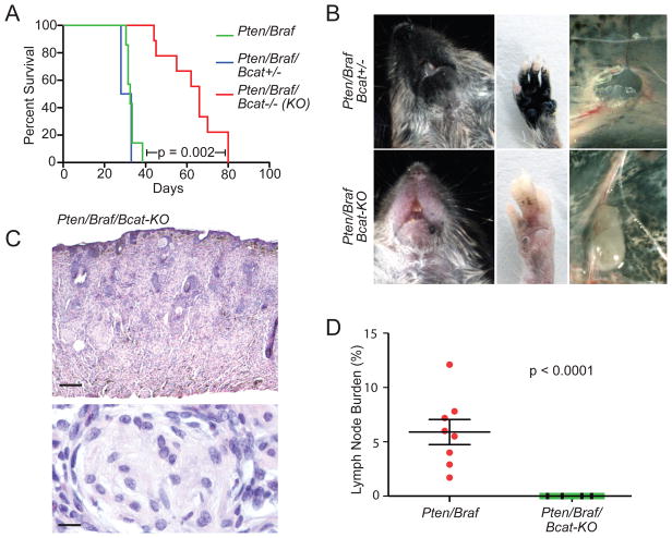
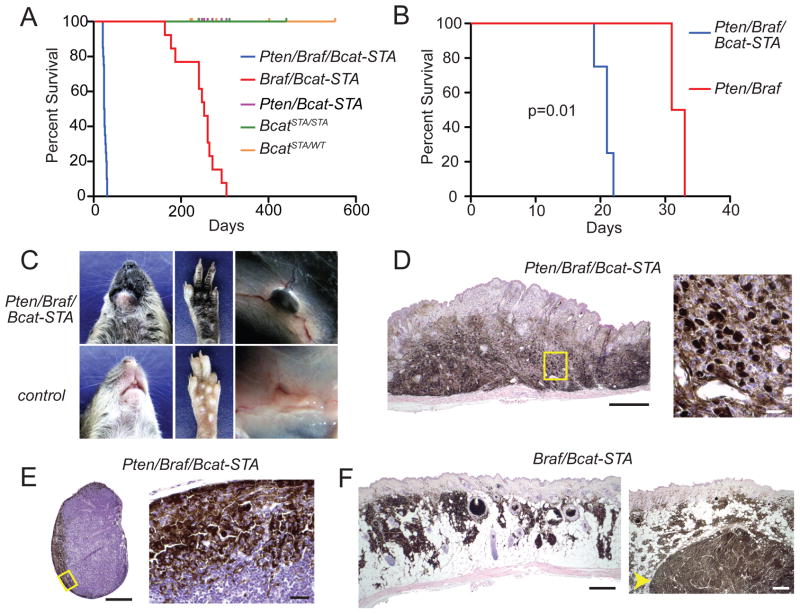
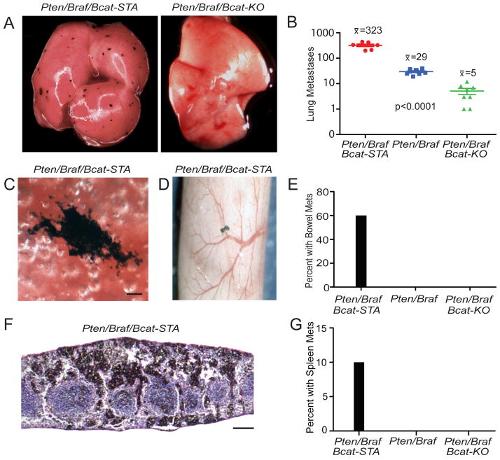
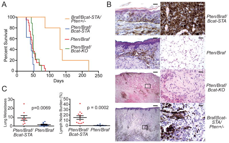
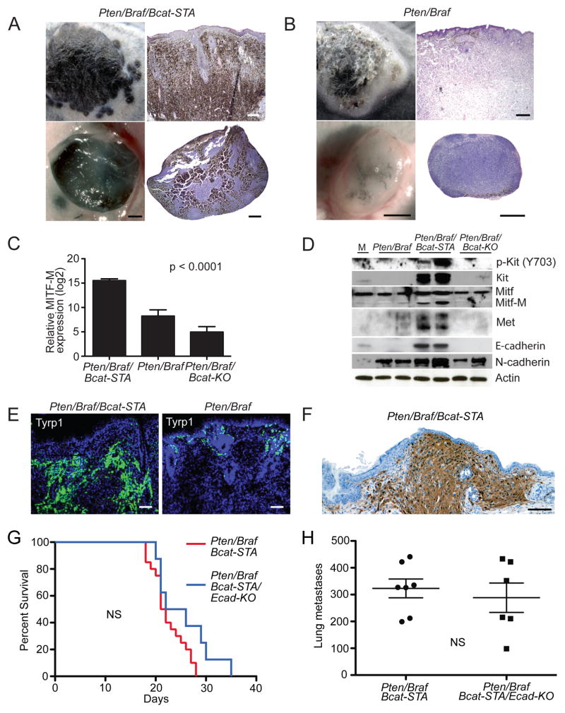
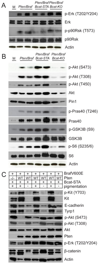
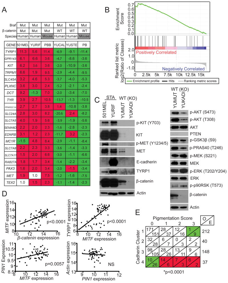
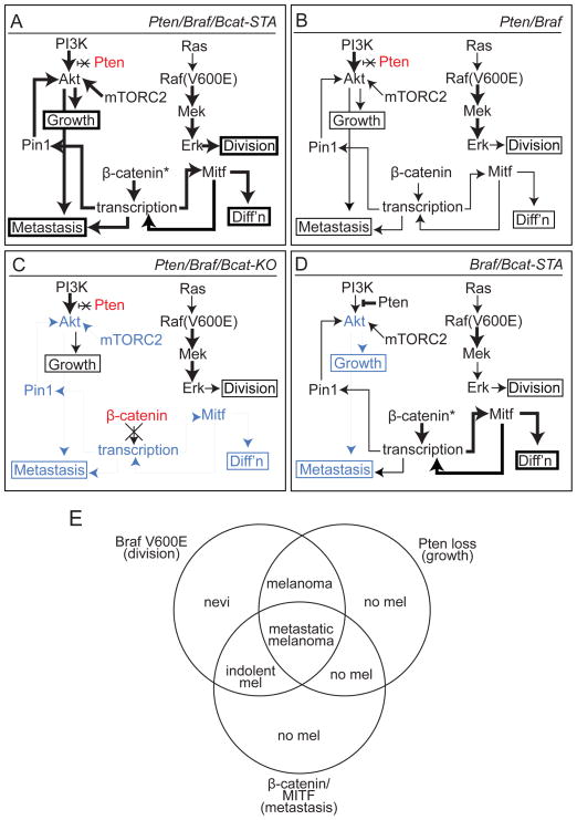
Malignant melanoma is characterized by frequent metastasis, however, specific changes that regulate this process have not been clearly delineated. Although it is well known that Wnt signaling is frequently dysregulated in melanoma, the functional implications of this observation are unclear. By modulating β-catenin levels in a mouse model of melanoma that is based on melanocyte-specific Pten loss and Braf(V600E) mutation, we demonstrate that β-catenin is a central mediator of melanoma metastasis to the lymph nodes and lungs. In addition to altering metastasis, β-catenin levels control tumor differentiation and regulate both MAPK/Erk and PI3K/Akt signaling. Highly metastatic tumors with β-catenin stabilization are very similar to a subset of human melanomas. Together these findings establish Wnt signaling as a metastasis regulator in melanoma.


Figures

Figure 1
Figure 1. β-catenin loss inhibits melanoma formation in Pten/Braf-driven melanomas
(A) Survival analysis of Pten/Braf cohorts after perinatal/generalized tumor induction. (B) Comparison of Pten/Braf/Bcat+/− (top) and Pten/Braf/Bcat-KO (bottom) tumor burden at 28 days of age. Metastasis to inguinal lymph node is also shown (right panels). (C) Hematoxylin and eosin (H&E) stained Pten/Braf/Bcat-KO flank tumor, scale 300 μM (top) and 10 μM (bottom). (D) Quantification of metastasis to the inguinal lymph nodes in Pten/Braf/Bcat-KO mice (n=13) compared to Pten/Braf mice (n=8). Error bars represent standard error of the mean (SEM). See also Figure S1.
Figure 2
Figure 2. β-catenin stabilization accelerates Pten/Braf-driven melanomagenesis
(A) Survival analysis of cohorts with β-catenin stabilization after perinatal tumor induction. (B) Survival analysis of Pten/Braf littermates, either with or without β-catenin stabilization, after perinatal tumor induction. (C) Tumor phenotype in Pten/Braf/Bcat-STA mice (top panels) compared to non-tumor prone mice (bottom panels) at 21 days of age. (D) H&E stained Pten/Braf/Bcat-STA flank tumor, scale 500 μM. Right panel: high power of outlined field, scale 20 μM. (E) H&E stained tumor-draining lymph node from a Pten/Braf/Bcat-STA mouse, scale 400 μM. High power of outlined field, scale 20 μM, right panel. (F) H&E stained nevi (left panel) and tumor (right panel, arrow) from a Braf/Bcat-STA mouse, scale 500 μM (left) and 500 μM (right). See also Table S1.
Figure 3
Figure 3. β-catenin signaling controls the ability of melanomas to metastasize to lung, bowel, and spleen
(A) Lungs from a perinatally treated Pten/Braf/Bcat-STA mouse at 27 days of age (left panel) and a perinatally treated Pten/Braf/Bcat-KO mouse at 56 days of age (right panel). (B) Comparison of number of lung metastases visible on the surface of the lung in perinatally treated litters. Error bars represent SEM. (C) Lung metastasis from a perinatally treated Pten/Braf/Bcat-STA mouse, scale 100 μM. (D) Bowel metastasis from a perinatally treated Pten/Braf/Bcat-STA mouse. (E) Comparison of number of bowel metastases in perinatally treated litters. (F) H&E stained spleen metastasis from a perinatally treated Pten/Braf/Bcat-STA mouse, scale 200 μM. (G) Comparison of number of spleen metastases in perinatally treated litters. See also Figure S2.
Figure 4
Figure 4. Locally induced melanomas are highly metastatic and can form lethal metastases
(A) Survival analysis of cohorts after localized tumor induction. Mice were euthanized when tumors reached 1 cm. (B) H&E stained sections of locally induced tumors from a Pten/Braf/Bcat-STA mouse (top panels, scale 20 μM left, 20 μM right), a Pten/Braf mouse (upper middle panels, scale 100 μM left, 50 μM right), a Pten/Braf/Bcat-KO mouse (lower middle panels, scale 400 μM left, 50 μM right), and a Braf/Bcat-STA/Pten+/− mouse (bottom panels, scale 400 μM left, 50 μM right). (C) Quantification of lung (left panel) and lymph node (right panel) metastasis in mice with focally induced melanomas. Error bars represent SEM. See also Figure S3 and Table S2.
Figure 5
Figure 5. Enhanced metastasis in Pten/Braf/Bcat-STA melanomas is accompanied by an increase in melanocytic differentiation markers
(A) Locally induced flank melanoma (upper left panel) with H&E (upper right panel, scale 400 μM) and draining lymph node (lower left panel, scale 500 μM) with H&E (lower right panel, scale 500 μM) in a Pten/Braf/Bcat-STA mouse. (B) Locally induced flank melanoma (upper left panel) with H&E (upper right panel, scale 400 μM) and draining lymph node (lower left panel, scale 500 μM) with H&E (lower right panel, scale 500 μM) in a Pten/Braf mouse.. (C) Mean expression of Mitf-M determined by qRT-PCR from Pten/Braf/Bcat-STA, Pten/Braf, and Pten/Braf/Bcat-KO melanomas (n=4 per genotype). Expression levels normalized to GAPDH control. (D) Western blots using protein lysates prepared directly from uncultured flank melanomas. M: normal cultured melanocytes. (E) Tyrp1 immunofluorescence of locally induced Pten/Braf/Bcat-STA (left panel) and Pten/Braf (right panel) flank melanomas, scale 200 μM. (F) S100 immunohistochemistry of a locally induced Pten/Braf/Bcat-STA melanoma from an albino mouse, scale 100 μM. (G) Survival analysis of Pten/Braf/Bcat-STA mice with or without E-cadherin inactivation after perinatal tumor induction, NS: not statistically significantly different. (H) Quantification of lung metastases in perinatally induced Pten/Braf/Bcat-STA mice with or without E-cadherin inactivation. Error bars represent SEM. See also Figure S4 and Table S3.
Figure 6
Figure 6. β-catenin stabilization is associated with hyperactivation of PI3K/Akt and MAPK/Erk signaling
(A–C) Western blots using protein lysates prepared from uncultured flank melanomas. M: Normal cultured melanocytes. See also Figure S5.
Figure 7
Figure 7. Pten/Braf/Bcat-STA murine melanomas faithfully recapitulate human melanomas
(A) Fold-change in expression of individual transcripts relative to mean for each transcript compared between human and murine melanomas. PBB: Pten/Braf/Bcat-STA, PB: Pten/Braf. (B) GSEA was performed on human melanomas with β-catenin stabilizing mutation using a subset of differentially regulated transcripts identified using murine melanomas, p<0.001. (C) Western blot analysis using protein lysates prepared from human melanoma cell lines. β-catenin status indicated for each sample. (D) Regression analysis comparing MITF, TYRP1, and PIN1 expression in a large human melanoma expression data set. (E) Contingency table of pigmentation status in human melanomas based on cadherin clusters (Kreizenbeck et al. 2008). O: observed, E: expected. Green: less than expected by chance, red: greater than expected by chance. See also Figure S6 and Table S4.
Figure 8
Figure 8. Signaling and phenotypic overview of murine melanoma models
(A) Pten/Braf/Bcat-STA melanomas are characterized by rapid growth, high degree of melanocytic differentiation, and extensive metastasis. These phenotypes are associated with increased MAPK/Erk and PI3K/Akt signaling as well as elevated Mitf and Pin1 levels. Bold text: relatively increased levels and/or activity compared to other models, blue shading: relatively decreased compared to other models, red shading: genetically inactivated, *: stabilized. (B) Pten/Braf melanomas exhibit rapid tumor growth, but only intermediate melanocytic differentiation and metastasis. Signaling pathway activation, Mitf, and Pin1 are intermediate in this model. (C) Pten/Braf/Bcat-KO melanomas exhibit tumor growth, but significantly reduced melanocytic differentiation and almost no metastasis. Inactivation of β-catenin in this model is associated with reduced PI3K/Akt signaling and Mitf levels. (D) Braf/Bcat-STA melanomas are characterized by long latency and slow tumor growth, as well as lack of metastases. These tumors are differentiated and heavily pigmented due to high Mitf levels, but show reduced PI3K/Akt pathway activation. (E) A three-pathway synergy is observed when MAPK/Erk, PI3K/Akt, and β-catenin/Mitf signaling pathways are simultaneously activated in melanoma. If any of these three changes is not present, the metastatic melanoma phenotype is abrogated.

References

    1. Bedrosian I, Faries MB, Guerry D, 4th, Elenitsas R, Schuchter L, Mick R, Spitz FR, Bucky LP, Alavi A, Elder DE, et al. Incidence of sentinel node metastasis in patients with thin primary melanoma (< or = 1mm) with vertical growth phase. Ann Surg Oncol. 2000;7:262–267. - PubMed
    1. Bennett DC, Dexter TJ, Ormerod EJ, Hart IR. Increased experimental metastatic capability of a murine melanoma following induction of differentiation. Cancer Res. 1986;46:3239–3244. - PubMed
    1. Boussadia O, Kutsch S, Hierholzer A, Delmas V, Kemler R. E-cadherin is a survival factor for the lactating mouse mammary gland. Mech Dev. 2002;115:53–62. - PubMed
    1. Brault V, Moore R, Kutsch S, Ishibashi M, Rowitch DH, McMahon AP, Sommer L, Boussadia O, Kemler R. Inactivation of the β-catenin gene by Wnt1-Cre-mediated deletion results in dramatic brain malformation and failure of craniofacial development. Development. 2001;128:1253–1264. - PubMed
    1. Chapman PB, Hauschild A, Robert C, Haanen JB, Ascierto P, Larkin J, Dummer R, Garbe C, Testori A, Maio M, et al. Improved survival with vemurafenib in melanoma with BRAF V600E mutation. N Engl J Med. 2011;364:2507–2516. - PMC - PubMed

Publication types

MeSH terms

Associated data