. 2011 Dec 13;20(6):768-80.
doi: 10.1016/j.ccr.2011.10.016.

WT1 mutants reveal SRPK1 to be a downstream angiogenesis target by altering VEGF splicing

Affiliations

WT1 mutants reveal SRPK1 to be a downstream angiogenesis target by altering VEGF splicing

Elianna M Amin et al. Cancer Cell. .

Abstract

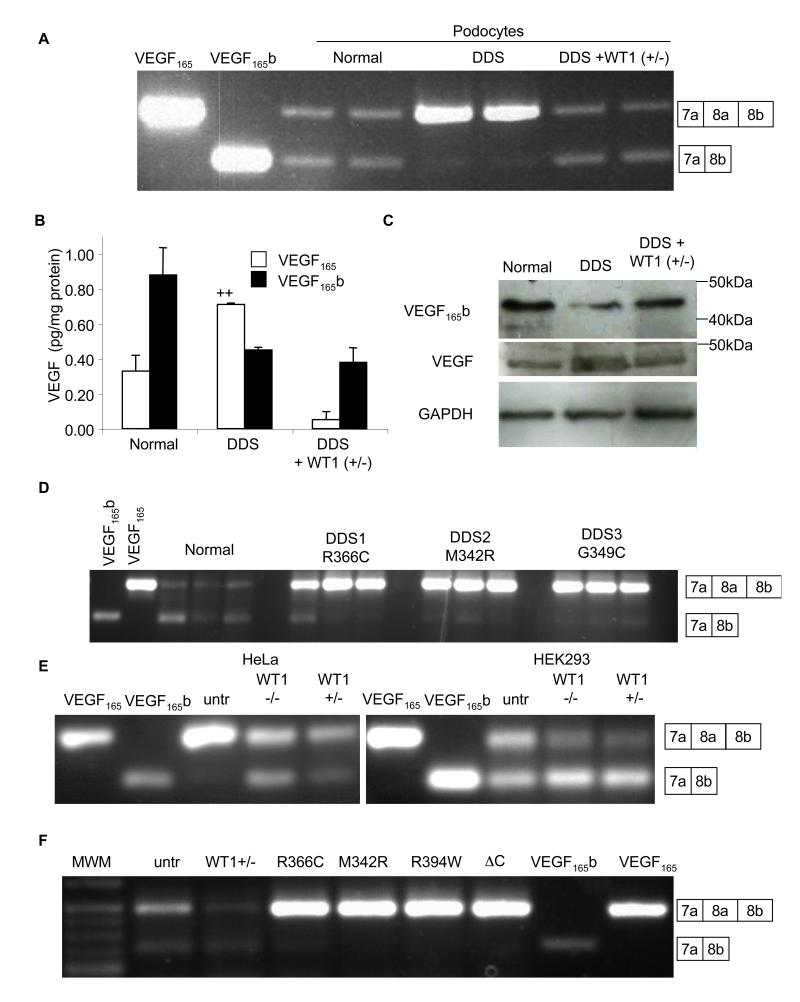
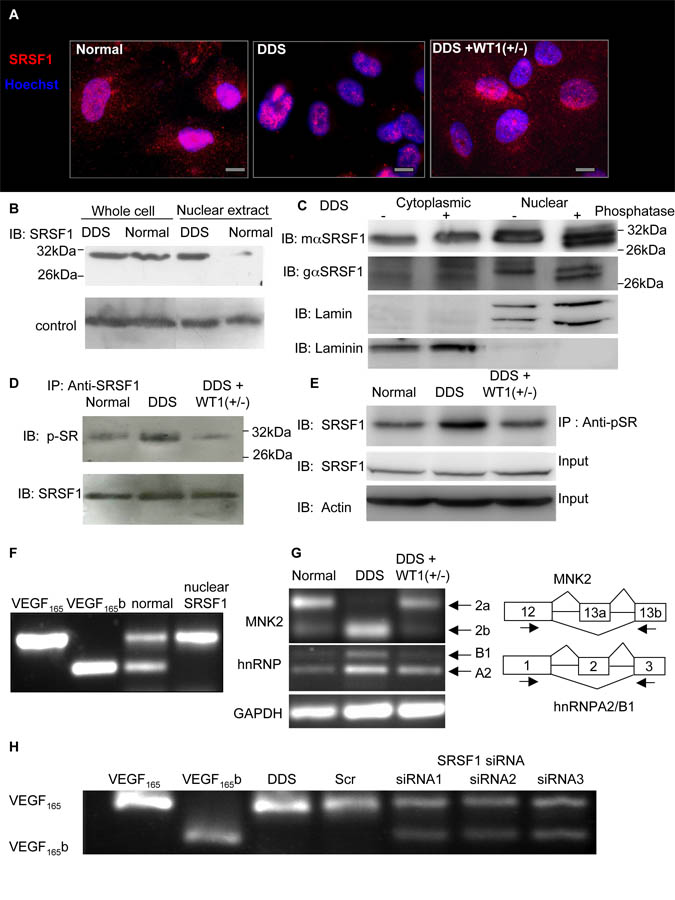
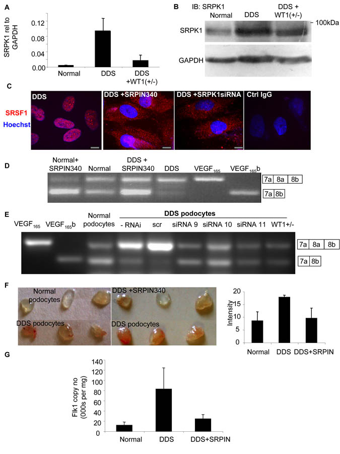
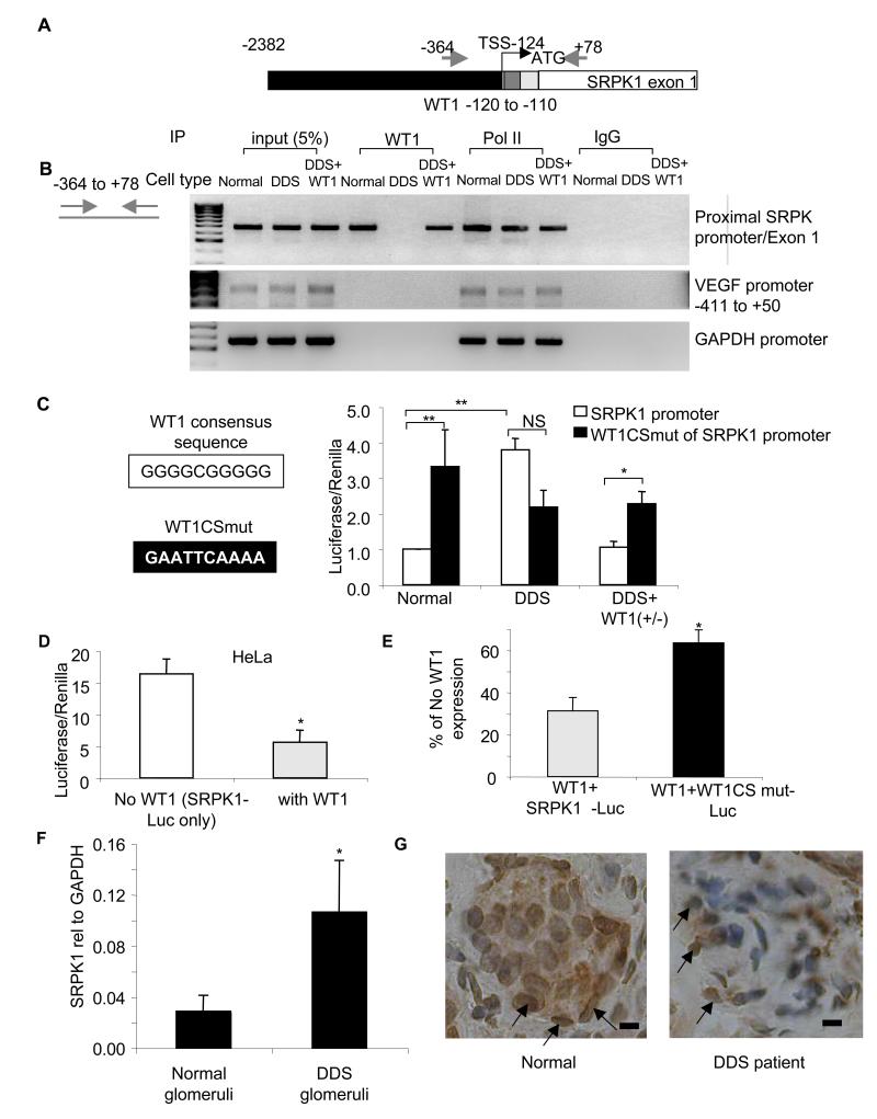
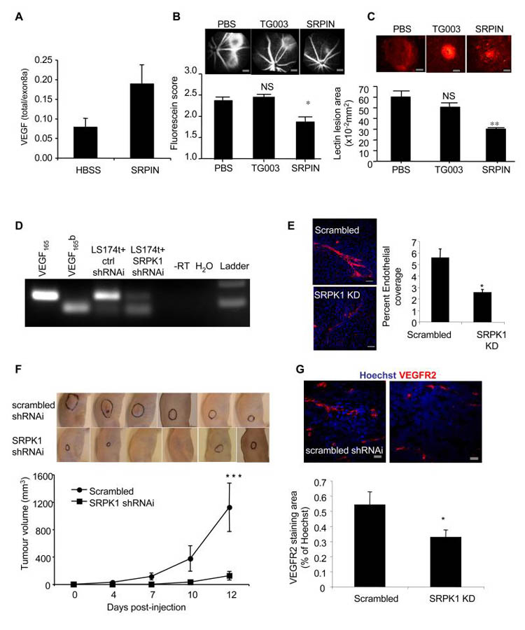
Angiogenesis is regulated by the balance of proangiogenic VEGF(165) and antiangiogenic VEGF(165)b splice isoforms. Mutations in WT1, the Wilms' tumor suppressor gene, suppress VEGF(165)b and cause abnormal gonadogenesis, renal failure, and Wilms' tumors. In WT1 mutant cells, reduced VEGF(165)b was due to lack of WT1-mediated transcriptional repression of the splicing-factor kinase SRPK1. WT1 bound to the SRPK1 promoter, and repressed expression through a specific WT1 binding site. In WT1 mutant cells SRPK1-mediated hyperphosphorylation of the oncogenic RNA binding protein SRSF1 regulated splicing of VEGF and rendered WT1 mutant cells proangiogenic. Altered VEGF splicing was reversed by wild-type WT1, knockdown of SRSF1, or SRPK1 and inhibition of SRPK1, which prevented in vitro and in vivo angiogenesis and associated tumor growth.


Figures

Figure 1
Figure 1. Wild type WT1 induces anti-angiogenic VEGF165b expression
A. RT-PCR of mRNA extracted from normal or DDS podocytes using primers that detect both proximal and distal splice isoforms of VEGF. VEGF165 and VEGF165b cDNAs are used as positive controls. Transfection of DDS podocytes with WT1 (+exon 5/−KTS) restored VEGF165b splicing. Boxes indicate exon usage represented by the bands. B. ELISAs for VEGF165b or VEGF165 protein using a pan-VEGF capture antibody and specific detection antibodies. VEGF165 was calculated from the difference between pan-VEGF and VEGF165b. C. Immunoblot of protein extracted from normal, DDS podocytes, or DDS cells transfected with wild type WT1(+/−) using antibodies to VEGF165b, total VEGF or GAPDH. D. RT-PCR of podocyte cell lines (three replicates) from three different DDS patients with different WT1 mutations show reduced VEGF165b expression. E. RT-PCR of mRNA from HeLa and HEK293 cells show expression of VEGF165b in HEK293 but not HeLa cells (untr=untransfected), but both cell types have increased VEGF165b expression in the presence of WT1 (−/−), and WT1 (+/−). F. Transfection of HEK293 cells with plasmids containing DDS-causing WT1 mutations abolished VEGF165b expression. Wild type WT1(+/−) over-expression increases distal splicing relative to proximally spliced isoforms. Bar charts are mean±SEM. See also figure S1.
Figure 2
Figure 2. Mutant WT1 induces phosphorylation and nuclear localisation of SRSF1, which regulates VEGF splicing
A. SRSF1 is absent from the cytoplasm when WT1 is mutated. Immunofluorescence staining of SRSF1 (red) of normal, DDS (R366C) and WT1 rescued DDS (DDS+WT1(+/−)) podocytes. Nuclei counterstained blue. Scale bar = 10μm. B. SRSF1 is more nuclearly localised in DDS than in normal podocytes. Protein was extracted from whole cells or from nuclear extracts of podocytes and subjected to immunoblotting for SRSF1. C. Nuclear SRSF1 is phosphorylated. Protein was extracted from flasks of DDS podocytes either as a cytoplasmic or a nuclear extract, and half treated with phosphatase and subjected to high resolution SDS PAGE and immunoblotting using mouse monoclonal (mαSRSF1), or goat polyclonal (gαSRSF1) antibodies to SRSF1, the cytoplasmic protein Laminin, and the nuclear protein Lamin. D SRSF1 is hyperphosphorylated in DDS. Protein was extracted from podocytes, immunoprecipitated with an SRSF1 antibody and immunoblotted with a panphospho SR antibody (top) or SRSF1 antibody (bottom). E. Protein was extracted from podocytes, and half immunoprecipitated with an anti-phosphoSR protein antibody. Both the IP and the crude protein extract were immunoblotted with a SRSF1 antibody and Actin as a loading control. F. Nuclear localisation of SRSF1 inhibits distal splicing. Normal podocytes were transfected with a vector encoding SRSF1 that fails to shuttle into the cytoplasm (nuclear specific), and RNA extracted and amplified for VEGF expression. G. Additional SRSF1 targets are alternatively spliced in DDS podocytes. RNA from normal or DDS podocytes was subjected to RT-PCR using exon spanning primers for the MNK2 cDNA (top) or hnRNPA2/B1 (middle) or GAPDH (bottom). H. Knockdown of SRSF1 induced VEGF165b expression in DDS podocytes. DDS podocytes were transfected with scrambled siRNA (scr) or three different siRNAs to SRSF1 in the 3′UTR (siRNA1), in exons 2-3 (siRNA2) or exon 3 (siRNA3). See also figure S2
Figure 3
Figure 3. Increased SRPK1 expression increases SRSF1 nuclear localisation and VEGF proximal splicing
A. SRPK1 is upregulated in DDS podocytes. mRNA was extracted from normal, DDS or rescued podocytes and subjected to Q-PCR using primers specific for SRPK1. B. SRPK1 protein is upregulated in DDS podocytes. Immunoblot (IB) using SRPK1 antibody in protein extracted from DDS podocytes, normal podocytes, and wild type WT1(+/−) over-expressing podocytes. C. SRSF1 localisation is SRPK1 dependent. Immunofluorescence for SRSF1 (red). DDS cells were treated with 10μM of the SRPK1 inhibitor SRPIN340 or by knockdown of SRPK1 in DDS podocytes. Scale bar = 10μm D. SRPK1 inhibition switches splicing to VEGF165b. RT-PCR of mRNA extracted from normal or DDS podocytes untreated or treated with the SRPK1 inhibitor SRPIN340 using primers for VEGF165 (top band) or VEGF165b (lower band). E. Knockdown of SRPK1 switches VEGF splicing. RT-PCR of RNA extracted from DDS podocytes transfected with three different siRNAs to SRPK1, or scrambled siRNA (scr). VEGF165b or VEGF165 cDNA is used as a positive control. F. SRPK1 inhibition is anti-angiogenic. Normal or DDS cells were suspended in matrigel with or without SRPIN340 and implanted into nude mice. Matrigel was excised and photographed. The intensity of red haemoglobin was measured. G. RNA was extracted from the matrigel plugs and endothelial content estimated from VEGFR2 expression level assessed by qRT-PCR. Bar charts are mean±SEM. See also figure S3.
Figure 4
Figure 4. WT1 represses SRPK1 expression by binding the SRPK1 promoter region
A. SRPK1 gene structure. Black=promoter region. Grey=WT1 binding site, silver=5′UTR, white=coding sequence of exon 1. Arrows=primer locations. Numbers are bp relative to start codon. B. WT1 binds the SRPK1 promoter. Nuclear extracts from podocytes were sheared to fragment DNA, immunoprecipitated with polymerase II antibody, human IgG or a WT1 antibody and subjected to PCR using primers to detect the region around the SRPK1 transcriptional start site (TSS), or the VEGF or GAPDH promoters. C. DDS mutations relieve SRPK1 repression. Podocytes were transfected with a SRPK1 promoter-luciferase reporter vector (white bars), or the same promoter with a 10base pair mutation of the WT1 consensus sequence (WT1CSmut, black bars). D. WT1 represses SRPK1 expression. HeLa cells were transfected with the wild type promoter reporter gene alone (open bars) or also with WT1 (silver bars), and bioluminescence measured. E. The 10bp consensus sequence adjacent to the TSS is responsible for part of the repression . HeLa cells transfected with WT1 and the wild type SRPK1-promoter (silver bars) or WT1 and the mutated SRPK1-promoter (black bars). Luciferase/Renilla is expressed as a percentage of bioluminescence in the absence of WT1 (white bar, in D) F. Q-PCR of SRPK1 RNA from histologically normal kidney (n=5) or children with DDS (n=5, p<0.05). Bar charts are mean±SEM. G. Immunohistochemistry for SRSF1 of kidney biopsies of DDS (right) compared with normal (left). Podocytes shown by arrows. Scale bar = 15μm, See also figure S4.
Figure 5
Figure 5. SRPK1 inhibition is anti-angiogenic
A. Mice underwent laser photocoagulation and were treated with SRPIN340 (inhibits SRPK1, n=24), TG003 (inhibits Clk1/4, n=18) or with vehicle (n=24). 2-3 days later six HBSS treated and six SRPIN340 treated mice were killed, eyes enucleated and RNA extracted and subjected to RT-qPCR for total VEGF (primers in exon 3) or exon 8a containing VEGF (e.g. VEGF164). B. The remaining mice were treated again seven days later with the compounds and then after another seven days subjected to fluorescein angiography. Lesion size was scored blind. Scale bar =300μm C. These mice were then killed and the retinal membranes flat mounted and stained for lectin to identify choroidal angiogenesis (red). The size of the lesions was measured. **=p<0.01, *=p<0.05 compared with PBS, ANOVA. Scale bar =100μm D. LS174t colon cancer cells were infected with lentivirus containing SRPK1-shRNAi or scrambled siRNA (ctrl) and selected with puromycin. Stable cell lines were subjected to RT-PCR for VEGF165b and VEGF165 levels. E. Knockdown of SRPK1 reduces the angiogenic potential of media. Cells were infected with SRPK1 shRNAi and the media collected. Media was used in an in vitro co-culture assay of endothelial cells and fibroblasts, and cells stained for CD31 (red, endothelial) and Hoechst (nuclei). Spread of endothelial cells was determined by measuring the ratio of area covered by CD31 staining relative to Hoechst staining. **=p<0.01. Scale bar = 200μm. F. Tumour cells were implanted into nude mice and tumours allowed to grow for 12 days. Tumours outlined in black. Tumour diameters were measured by callipers. ***=p<0.001 by two way ANOVA. G. Tumours were excised and stained for VEGFR2 (red) and Hoechst (blue) and vessel density determined by VEGFR2 staining relative to Hoechst. , *=p<0.05 compared with scrambled siRNA, t test. Scale bar =20μm. Bar charts are mean±SEM. See also figure S5.

Comment in

References

    1. Artac RA, McFee RM, Smith RA, Baltes-Breitwisch MM, Clopton DT, Cupp AS. Neutralization of vascular endothelial growth factor antiangiogenic isoforms is more effective than treatment with proangiogenic isoforms in stimulating vascular development and follicle progression in the perinatal rat ovary. Biol Reprod. 2009;81:978–988. - PMC - PubMed
    1. Ballmer-Hofer K, Andersson AE, Ratcliffe LE, Berger P. Neuropilin-1 promotes VEGFR-2 trafficking through Rab11 vesicles thereby specifying signal output. Blood. 2011 - PubMed
    1. Baltes-Breitwisch MM, Artac RA, Bott RC, McFee RM, Kerl JG, Clopton DT, Cupp AS. Neutralization of vascular endothelial growth factor antiangiogenic isoforms or administration of proangiogenic isoforms stimulates vascular development in the rat testis. Reproduction. 2010;140:319–329. - PMC - PubMed
    1. Bates DO, Cui TG, Doughty JM, Winkler M, Sugiono M, Shields JD, Peat D, Gillatt D, Harper SJ. VEGF165b, an inhibitory splice variant of vascular endothelial growth factor, is down-regulated in renal cell carcinoma. Cancer Res. 2002;62:4123–4131. - PubMed
    1. Blann AD, Li JL, Li C, Kumar S. Increased serum VEGF in 13 children with Wilms’ tumour falls after surgery but rising levels predict poor prognosis. Cancer Lett. 2001;173:183–186. - PubMed

Publication types

MeSH terms

LinkOut - more resources