Review
. 2012 Sep-Oct;1819(9-10):1035-54.
doi: 10.1016/j.bbagrm.2011.11.009. Epub 2011 Dec 7.

Mechanism of protein biosynthesis in mammalian mitochondria

Affiliations
Review

Mechanism of protein biosynthesis in mammalian mitochondria

Brooke E Christian et al. Biochim Biophys Acta. 2012 Sep-Oct.

Abstract

Protein synthesis in mammalian mitochondria produces 13 proteins that are essential subunits of the oxidative phosphorylation complexes. This review provides a detailed outline of each phase of mitochondrial translation including initiation, elongation, termination, and ribosome recycling. The roles of essential proteins involved in each phase are described. All of the products of mitochondrial protein synthesis in mammals are inserted into the inner membrane. Several proteins that may help bind ribosomes to the membrane during translation are described, although much remains to be learned about this process. Mutations in mitochondrial or nuclear genes encoding components of the translation system often lead to severe deficiencies in oxidative phosphorylation, and a summary of these mutations is provided. This article is part of a Special Issue entitled: Mitochondrial Gene Expression.


Figures

Figure 1
Figure 1
Secondary structures of three mitochondrial mRNAs. RNA SHAPE chemistry was used to analyze the structures of the 5′ ends of the mitochondrial mRNAs and indicated that most are largely unstructured [26]. The secondary structures of the NADH dehydrogenase subunits 3 (MTND3) and 5 (MTND5) mRNAs, as well as the cytochrome b (CytB) mRNA are shown. The free energy values for each stem-loop structure are indicated adjacent to the stem-loop.
Figure 2
Figure 2
Model for the initiation phase of mitochondrial translation. In the current model for the initiation of protein synthesis, mitochondrial initiation factor 3 (IF3mt) actively dissociates 55S ribosomes, forming a transient [IF3mt:55S] complex (Step 1) and leading to the formation of an IF3mt:28S complex (Step 2). Mitochondrial initiation factor 2 (IF2mt) bound to GTP binds to the small subunit (Step 3), followed by the fMet-tRNA and mRNA (Step 4), although the exact order of binding is not clear. The mRNA feeds into the mRNA entrance gate and the 5′ end pauses at the P-site of the ribosome for inspection of its 5′ start codon. In the presence of the correct start codon and fMet-tRNA, the mRNA is locked in place by codon:anticodon interactions to form the 28S initiation complex. If fMet-tRNA binds in the absence of mRNA, or if the mRNA does not contain a proper start codon, the inspection step fails. The failed inspection causes the mRNA to continue sliding through the ribosome to exit. Once the 28S initiation complex has formed, the large subunit joins, and along with the hydrolysis of GTP to GDP, the initiation factors exit (Step 5) leaving a 55S:fMet-tRNA:mRNA complex that is ready for the elongation phase of protein synthesis.
Figure 3
Figure 3
Organization of initiation factor 2 (IF2). A. Domain alignment of E. coli, G. stearothermophilus, and M. thermoautotrophicum IF2, as well as mammalian IF2mt. B. Model for the 3-D structure of IF2mt, based on the cryo-EM map of IF2mt [63]. Domain 3 has been omitted from the structure, since cryo-EM images were not of sufficient resolution to obtain its coordinates, and no corresponding structures were available in the databases upon which to build a model. Domain IV is shown in blue, domain V is shown in purple, the insertion domain is shown in orange, domain VI-C1 is shown in green, and domain VI-C2 is shown in pink.
Figure 4
Figure 4
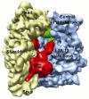
Binding of IF2mt to the E. coli ribosome. This image was taken from the cryo-EM structure of the 70S:mRNA:fMet-tRNA:IF2mt:GDPNP complex [63] and shows the orientation of IF2mt on the E. coli ribosome. The bacterial 30S subunit is shown in light yellow and the bacterial 50S subunit is shown in blue. Defining features of each subunit are labeled, including the head, shoulder and spur of the small subunit and the central protuberance and L7/L12 stalk base on the large subunit. IF2mt is shown in red, and the initiator tRNA is shown in green. The authors thank Dr. Rajendra Agrawal, Wadsworth Center, New York State Dept. of Health, Albany, NY for the cryo-EM image.
Figure 5
Figure 5
Organization of IF3mt. A. Domain organization of IF3mt. The regions of IF3mt with homology to bacterial IF3 are indicated. B. 3-D model of IF3mt based on the crystal structure of the N-terminal domain of G. stearothermophilus IF3 and the NMR structure of the C-terminal domain of mouse IF3mt [66]. The N-extension is shown using a dashed grey line and could not be modeled due to its predicted lack of structure. The N-domain is shown in yellow, the linker is shown in pink, and the C-domain is shown in blue. The C-extension is shown using a dashed green line and could not be modeled since no structure could be predicted for this region of IF3mt.
Figure 6
Figure 6
Interaction sites of IF3mt on the small ribosomal subunit. The structure of a “mock” mitochondrial 28S small subunit was prepared based on the crystal structure of the Thermus thermophilus 30S ribosomal subunit. The small subunit is shown with the interface side facing the reader and only homologous proteins and RNA segments present in the mitochondrial 28S subunit are shown. The ribosomal proteins that can cross-link to IF3mt are spaced-filled. Small subunit protein S5 is shown in orange, S9 in red, S10 in green, and S18 in blue.
Figure 7
Figure 7
Model for the elongation phase of mitochondrial translation. The tRNA containing the growing polypeptide chain is located in the P-site of the ribosome. EF-Tumt brings the aa-tRNA to the A-site of the ribosome (Step 1). In concert with the hydrolysis of GTP to GDP, EF-Tumt leaves the ribosome (Step 2). EF-Tsmt binds to EF-Tumt, displacing the GDP molecule and forming an EF-Tumt·EF-Tsmt complex (Step 3). A GTP molecule displaces EF-Tsmt, and an EF-Tumt:GTP complex is formed (Step 4) which can then bind another aa-tRNA reforming the ternary complex. The large ribosomal subunit catalyzes peptide bond formation and the growing polypeptide chain is transferred to the tRNA in the A-site of the ribosome (Step 5). EF-G1mt:GTP binds to the ribosome at the A-site (Step 6) and catalyzes translocation of the ribosome, moving the deacylated tRNA out of the P-site and the peptidyl-tRNA from the A-site to the P-site (Step 7). A new cycle of elongation can then begin.
Figure 8
Figure 8
Regions of EF-Tumt responsible for interacting with aa-tRNA. A. Alignment of EF-Tumt and E. coli EF-Tu domain II [106]. The amino acid residues of domain II that interact with the 3′ acceptor stem region of the aminoacyl-tRNA are indicated with a •, and the residues that interact with the 5′ end of the tRNA are indicated with a B. Interaction of EF-Tu with Cys-tRNA based on the crystal structure of T. aquaticus EF-Tu:GDPNP:E. coli Cys-tRNACys (PDB# 1B23). C. Close-up image of the box in B. Image is rotated approximately 90 degrees. The Cys-tRNACys is shown in gray, EF-Tu is shown in orange, and GDPNP is shown in purple. The residues of EF-Tu that contact the tRNA are shown in pink, the 5′ G of the tRNA is shown in blue and the A of the 3′ -CCA end of the tRNA is green. The 5′ phosphate group of the tRNA is indicated by the black arrow.
Figure 9
Figure 9
Crystal structure of the bovine EF-Tumt:EF-Tsmt complex. In the 3-D structure of the bovine EF-Tumt:EF-Tsmt complex (PDB coordinates 1XB2) [101], EF-Tumt is shown in orange and EF-Tsmt is in blue. The domains of each protein are labeled and the position of the insertion present in one isoform of EF-Tsmt is indicated by a red line.
Figure 10
Figure 10
Model for the termination and ribosome recycling phases of mitochondrial protein synthesis. As the termination codon (UAG here) enters the A-site of the ribosome, mtRF1a and GTP bind to the A-site (Step 1) and promote GTP-dependent hydrolysis and release of the polypeptide chain (Step 2). How mtRF1a is released from the ribosome is not known. RRF1mt binds to the A-site of the ribosome (Step 3) and is joined by RRF2mt (also termed EF-G2mt). These factors promote the dissociation of the ribosomal subunits and release of the deacylated tRNA and the mRNA (Step 4). Following release of RRF1mt and RRF2mt (Step 5), the ribosome begins another round of protein synthesis.
Figure 11
Figure 11
Interaction sites of the C-terminal tail of Oxa1L (Oxa1L-CTT) on the large ribosomal subunit. The 3-D structure of the T. thermophilus 50S ribosomal subunit (PDB 2WRL) is shown viewed from the solvent side. Regions of the rRNA that are not present in the mitochondrial 39S subunit have been removed. Mammalian homologs of bacterial ribosomal proteins predicted to interact with Oxa1L-CTT based on cross-linking studies are shown as space-filled with L13 (red spheres), L20 (green spheres) and L28 (blue spheres). The proteins thought to make up the traditional polypeptide exit tunnel (L22, L23, L24, and L29) are shown in orange spheres and do not cross-link to Oxa1L-CTT. Other large subunit ribosomal subunits are shown in black.

References

    1. Vogel F, Bornhovd C, Neupert W, Reichert AS. Dynamic subcompartmentalization of the mitochondrial inner membrane. J. Cell Biol. 2006;175:237–247. - PMC - PubMed
    1. Osawa S, Jukes T, Watanabe K, Muto A. Recent evidence for evolution of the genetic code. Micro. Rev. 1992;56:229–264. - PMC - PubMed
    1. Temperley R, Richter R, Dennerlein S, Lightowlers RN, Chrzanowska- Lightowlers ZM. Hungry codons promote frameshifting in human mitochondrial ribosomes. Science. 2010;327:301. - PubMed
    1. Taanman J-W. The mitochondrial genome: structure, transcription, translation and replication. Biochim. Biophys. Acta. 1999;1410:103–123. - PubMed
    1. O’Brien TW. The general occurrence of 55 S ribosomes in mammalian liver mitochondria. J. Biol. Chem. 1971;246:3409–3417. - PubMed

Publication types

MeSH terms

LinkOut - more resources