

Mitochondrial poly(A) polymerase and polyadenylation
- PMID: 22172994
- PMCID: PMC3307840
- DOI: 10.1016/j.bbagrm.2011.10.012
Mitochondrial poly(A) polymerase and polyadenylation
Abstract
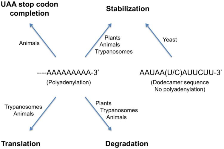
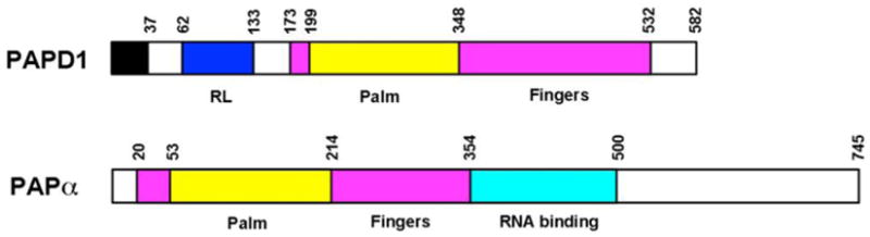
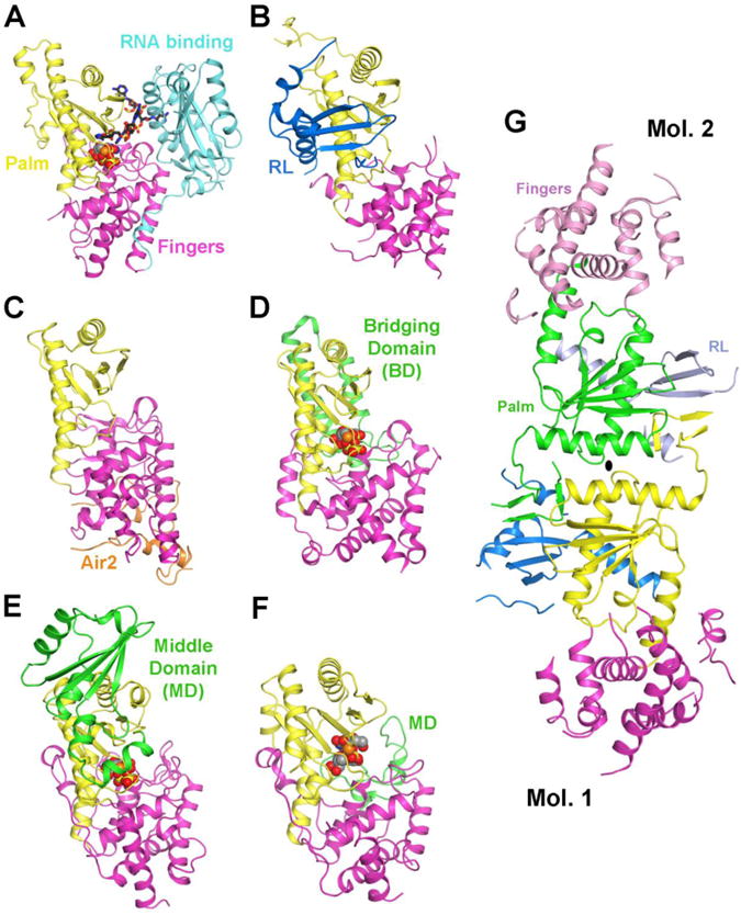
Polyadenylation of mitochondrial RNAs in higher eukaryotic organisms have diverse effects on their function and metabolism. Polyadenylation completes the UAA stop codon of a majority of mitochondrial mRNAs in mammals, regulates the translation of the mRNAs, and has diverse effects on their stability. In contrast, polyadenylation of most mitochondrial mRNAs in plants leads to their degradation, consistent with the bacterial origin of this organelle. PAPD1 (mtPAP, TUTase1), a noncanonical poly(A) polymerase (ncPAP), is responsible for producing the poly(A) tails in mammalian mitochondria. The crystal structure of human PAPD1 was reported recently, offering molecular insights into its catalysis. This article is part of a Special Issue entitled: Mitochondrial Gene Expression.
Copyright © 2011 Elsevier B.V. All rights reserved.
Figures
Similar articles
-
Helicase SUV3, polynucleotide phosphorylase, and mitochondrial polyadenylation polymerase form a transient complex to modulate mitochondrial mRNA polyadenylated tail lengths in response to energetic changes.J Biol Chem. 2014 Jun 13;289(24):16727-35. doi: 10.1074/jbc.M113.536540. Epub 2014 Apr 25. J Biol Chem. 2014. PMID: 24770417 Free PMC article.
-
Structural basis for dimerization and activity of human PAPD1, a noncanonical poly(A) polymerase.Mol Cell. 2011 Feb 4;41(3):311-20. doi: 10.1016/j.molcel.2011.01.013. Mol Cell. 2011. PMID: 21292163 Free PMC article.
-
LRPPRC/SLIRP suppresses PNPase-mediated mRNA decay and promotes polyadenylation in human mitochondria.Nucleic Acids Res. 2012 Sep;40(16):8033-47. doi: 10.1093/nar/gks506. Epub 2012 May 31. Nucleic Acids Res. 2012. PMID: 22661577 Free PMC article.
-
Polyadenylation-assisted RNA degradation processes in plants.Trends Plant Sci. 2009 Sep;14(9):497-504. doi: 10.1016/j.tplants.2009.06.007. Epub 2009 Aug 27. Trends Plant Sci. 2009. PMID: 19716749 Review.
-
Polyadenylation in mammalian mitochondria: insights from recent studies.Biochim Biophys Acta. 2008 Apr;1779(4):266-9. doi: 10.1016/j.bbagrm.2008.02.001. Epub 2008 Feb 14. Biochim Biophys Acta. 2008. PMID: 18312863 Review.
Cited by
-
Variation and evolution of polyadenylation profiles in sauropsid mitochondrial mRNAs as deduced from the high-throughput RNA sequencing.BMC Genomics. 2017 Aug 29;18(1):665. doi: 10.1186/s12864-017-4080-0. BMC Genomics. 2017. PMID: 28851277 Free PMC article.
-
In Planta Determination of the mRNA-Binding Proteome of Arabidopsis Etiolated Seedlings.Plant Cell. 2016 Oct;28(10):2435-2452. doi: 10.1105/tpc.16.00562. Epub 2016 Oct 11. Plant Cell. 2016. PMID: 27729395 Free PMC article.
-
Exploration of CCA-added RNAs revealed the expression of mitochondrial non-coding RNAs regulated by CCA-adding enzyme.RNA Biol. 2019 Dec;16(12):1817-1825. doi: 10.1080/15476286.2019.1664885. Epub 2019 Sep 12. RNA Biol. 2019. PMID: 31512554 Free PMC article.
-
A Uniquely Complex Mitochondrial Proteome from Euglena gracilis.Mol Biol Evol. 2020 Aug 1;37(8):2173-2191. doi: 10.1093/molbev/msaa061. Mol Biol Evol. 2020. PMID: 32159766 Free PMC article.
-
Infection by a Giant Virus (AaV) Induces Widespread Physiological Reprogramming in Aureococcus anophagefferens CCMP1984 - A Harmful Bloom Algae.Front Microbiol. 2018 Apr 19;9:752. doi: 10.3389/fmicb.2018.00752. eCollection 2018. Front Microbiol. 2018. PMID: 29725322 Free PMC article.
References
Publication types
MeSH terms
Substances
Grants and funding
LinkOut - more resources
Full Text Sources
Other Literature Sources
