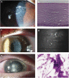
Microsporidia and Acanthamoeba: the role of emerging corneal pathogens
- PMID: 22173072
- PMCID: PMC3272212
- DOI: 10.1038/eye.2011.315
Microsporidia and Acanthamoeba: the role of emerging corneal pathogens
Abstract
Parasitic organisms are increasingly recognized as human corneal pathogens. A notable increase in both well-defined Acanthamoeba keratitis and a more dramatic increase in reported cases of Microsporidia keratitis have suggested significant outbreaks of parasitic keratitis around the world. Historical and contemporary baselines as well as a familiar associated clinical presentation reinforce the significant outbreak of Acanthamoeba keratitis in the United States. The remarkable rise in cases of Microsporidia keratitis, however, lacks these established baselines and, further, describes a disease that is inconsistent with previous definitions of disease. While a well-defined, abrupt increase strongly suggests temporally related risk factors, most likely environmental, involved in the Acanthamoeba outbreak, the rise in Microsporidia keratitis suggests that increased awareness and improved diagnostic acumen are a significant factor in case ascertainment. Regardless, recent evidence indicates that both parasitic diseases are likely underreported in various forms of infectious keratitis, which may have unrecognized but significant implications in the pathogenesis of both primary protozoal and polymicrobial keratitis. Further understanding of the incidence and interaction of these organisms with current therapeutic regimens and more commonly recognized pathogens should significantly improve diagnosis and alter clinical outcomes.
Figures

Similar articles
-
A DNA dot hybridization model for molecular diagnosis of parasitic keratitis.Mol Vis. 2017 Aug 24;23:614-623. eCollection 2017. Mol Vis. 2017. PMID: 28867932 Free PMC article.
-
American Academy of Optometry Microbial Keratitis Think Tank.Optom Vis Sci. 2021 Mar 1;98(3):182-198. doi: 10.1097/OPX.0000000000001664. Optom Vis Sci. 2021. PMID: 33771951 Free PMC article.
-
Diagnostic Evaluation of Co-Occurrence of Acanthamoeba and Fungi in Keratitis: A Preliminary Report.Cornea. 2018 Feb;37(2):227-234. doi: 10.1097/ICO.0000000000001441. Cornea. 2018. PMID: 29111995
-
Microsporidial keratitis: need for increased awareness.Surv Ophthalmol. 2011 Jan-Feb;56(1):1-22. doi: 10.1016/j.survophthal.2010.03.006. Epub 2010 Nov 11. Surv Ophthalmol. 2011. PMID: 21071051 Review.
-
PCR-based diagnosis and clinical insights into parasitic keratitis.J Microbiol Immunol Infect. 2025 Jun;58(3):363-367. doi: 10.1016/j.jmii.2025.01.002. Epub 2025 Jan 22. J Microbiol Immunol Infect. 2025. PMID: 39870545
Cited by
-
Recurrent interface abscess secondary to Acanthamoeba keratitis treated by deep anterior lamellar keratoplasty.Int J Ophthalmol. 2012;5(6):774-5. doi: 10.3980/j.issn.2222-3959.2012.06.22. Epub 2012 Dec 18. Int J Ophthalmol. 2012. PMID: 23275916 Free PMC article. No abstract available.
-
A DNA dot hybridization model for molecular diagnosis of parasitic keratitis.Mol Vis. 2017 Aug 24;23:614-623. eCollection 2017. Mol Vis. 2017. PMID: 28867932 Free PMC article.
-
Results of case-control studies support the association between contact lens use and Acanthamoeba keratitis.Clin Ophthalmol. 2013;7:991-4. doi: 10.2147/OPTH.S43471. Epub 2013 May 28. Clin Ophthalmol. 2013. PMID: 23761962 Free PMC article.
-
Effect of low concentrations of benzalkonium chloride on acanthamoebal survival and its potential impact on empirical therapy of infectious keratitis.JAMA Ophthalmol. 2013 May;131(5):595-600. doi: 10.1001/jamaophthalmol.2013.1644. JAMA Ophthalmol. 2013. PMID: 23519403 Free PMC article.
-
Parasitic keratitis - An under-reported entity.Trop Parasitol. 2020 Jan-Jun;10(1):12-17. doi: 10.4103/tp.TP_84_19. Epub 2020 May 20. Trop Parasitol. 2020. PMID: 32775286 Free PMC article.
References
-
- Pearlman E, Gillette-Ferguson I. Onchocerca volvulus, Wolbachia and river blindness. Chem Immunol Allergy. 2007;92:254–265. - PubMed
-
- Lam S. Keratitis caused by leishmaniasis and trypanosomiasis. Ophthalmol Clin N Am. 1994;7 (4:635–639.
-
- Thebpatiphat N, Hammersmith KM, Rocha FN, Rapuano CJ, Ayres BD, Laibson PR, et al. Acanthamoeba keratitis: a parasite on the rise. Cornea. 2007;26 (6:701–706. - PubMed
MeSH terms
Grants and funding
LinkOut - more resources
Full Text Sources

