. 2012 Jun;26(6):1218-27.
doi: 10.1038/leu.2011.358. Epub 2011 Dec 16.

Digital gene expression profiling of primary acute lymphoblastic leukemia cells

Affiliations
Free PMC article

Digital gene expression profiling of primary acute lymphoblastic leukemia cells

J Nordlund et al. Leukemia. 2012 Jun.
Free PMC article

Abstract

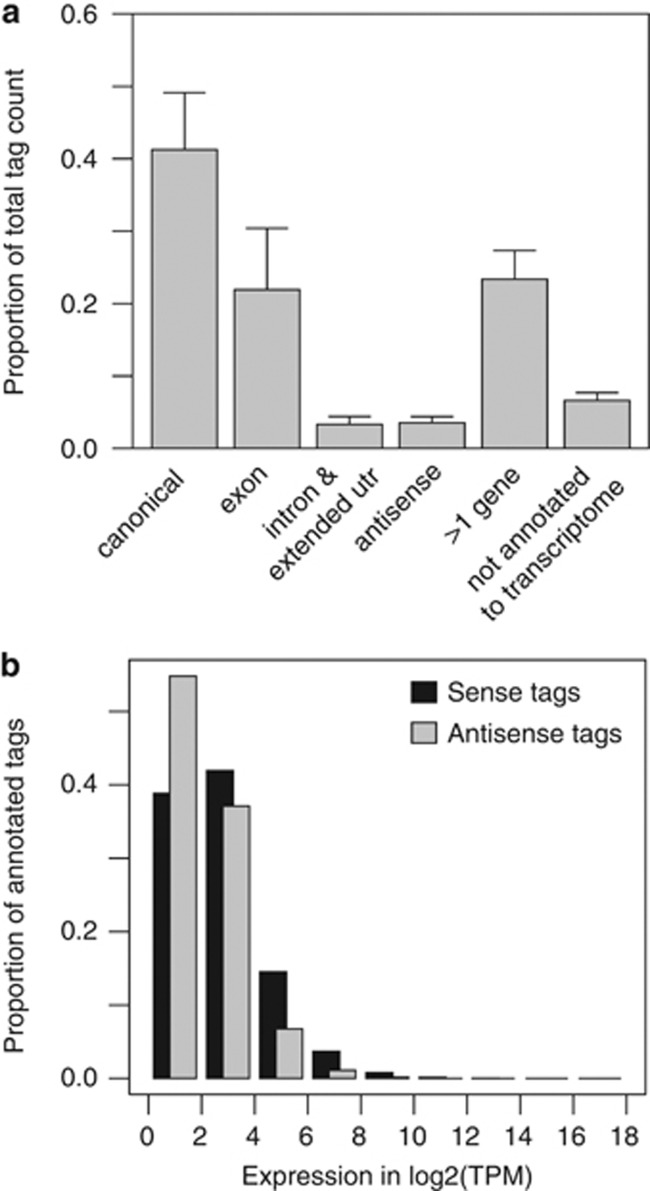
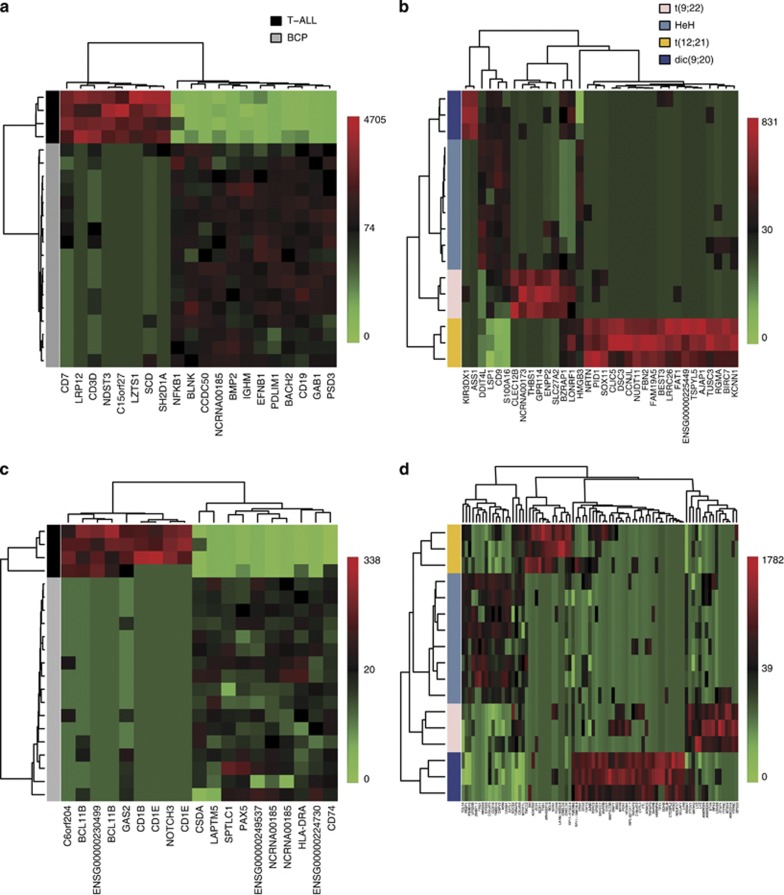
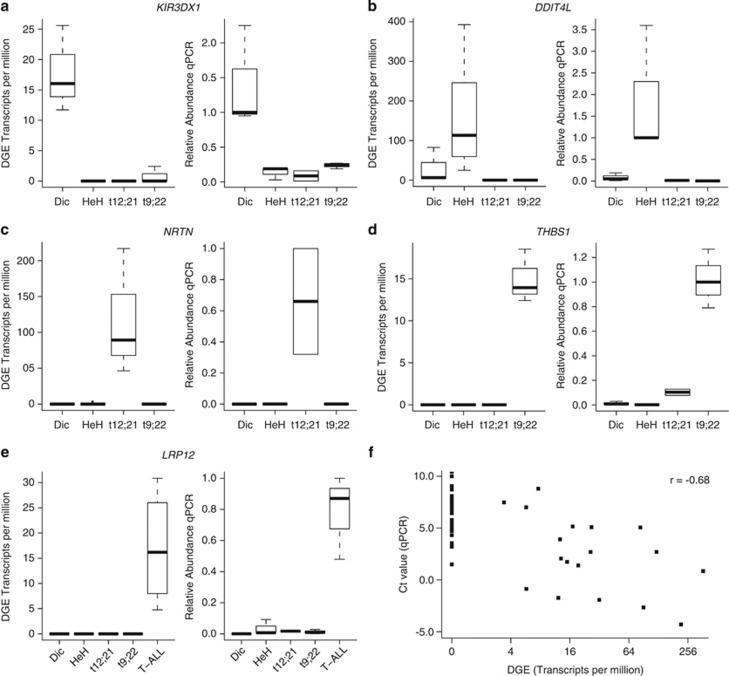
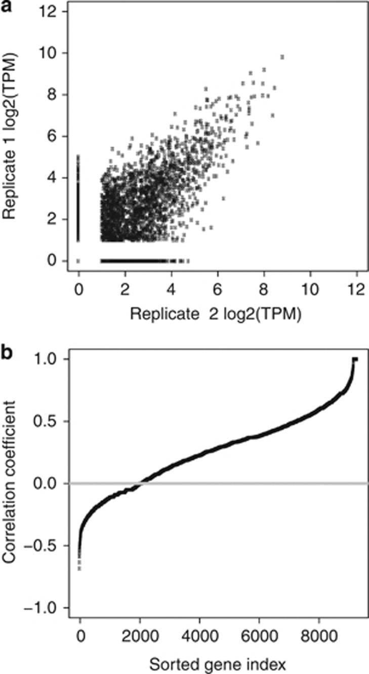
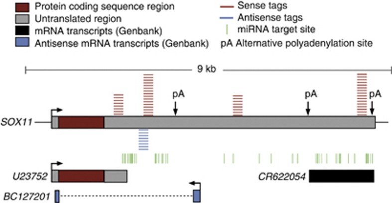
We determined the genome-wide digital gene expression (DGE) profiles of primary acute lymphoblastic leukemia (ALL) cells from 21 patients taking advantage of 'second-generation' sequencing technology. Patients included in this study represent four cytogenetically distinct subtypes of B-cell precursor (BCP) ALL and T-cell lineage ALL (T-ALL). The robustness of DGE combined with supervised classification by nearest shrunken centroids (NSC) was validated experimentally and by comparison with published expression data for large sets of ALL samples. Genes that were differentially expressed between BCP ALL subtypes were enriched to distinct signaling pathways with dic(9;20) enriched to TP53 signaling, t(9;22) to interferon signaling, as well as high hyperdiploidy and t(12;21) to apoptosis signaling. We also observed antisense tags expressed from the non-coding strand of ~50% of annotated genes, many of which were expressed in a subtype-specific pattern. Antisense tags from 17 gene regions unambiguously discriminated between the BCP ALL and T-ALL subtypes, and antisense tags from 76 gene regions discriminated between the 4 BCP subtypes. We observed a significant overlap of gene regions with alternative polyadenylation and antisense transcription (P<1 × 10(-15)). Our study using DGE profiling provided new insights into the RNA expression patterns in ALL cells.


Figures

Figure 1
Figure 1
Distribution between gene locations and abundance of annotated tags. To annotate the sequenced tags, we created a database of all possible 17-bp tag sequences next to an NlaIII site from the Ensembl transcriptome database. (a) The bars show the proportion of the total number of tags by location in gene regions. The canonical location refers to the tag originating from the most 3′ NlaIII site in a gene, to which 41% mapped. Overall, 22% of the tags were mapped to exons, which may represent transcript isoforms that are not listed in Ensembl. In all, 3.4% of the tags, which presumably originate from unprocessed pre-mRNA transcripts, mis-spliced transcripts, or unannotated exons, were annotated to intronic gene sequences. Overall, 3.6% of the sequenced tags mapped in an antisense orientation to gene regions in the Ensembl database. The remaining tags had multiple annotations (23%) or were not found in our database of possible tag sequences (7%). No significant difference in the distribution between gene locations was observed for the annotated tags between the ALL subtypes (data not shown). (b) The bars show the proportion of annotated tags at different bins of expression levels. The expression levels are in tags per million (TPM) on a log2- transformed scale on the horizontal axis. Black bars indicate tags annotated to genes in the sense direction and light gray bars indicate tags annotated to genes in the antisense direction.
Figure 2
Figure 2
Hierarchical clustering of ALL samples according to gene regions defined by nearest shrunken centroid (NSC) classification based on DGE data from sense and antisense transcripts. (a) Heatmap of the expression levels of the 20 genes defined by the NSC classifier for discriminating between the samples of BCP and T-ALL subtypes. These genes are listed in Supplementary Table S2. (b) Heatmap of the expression levels of 34 genes defined by the NSC classifier for discriminating between the 4 BCP subtypes. The 34 genes are listed in Supplementary Table S3. (c) Heatmap of the expression levels of 19 antisense expressed tags defined by the NSC classifier for discriminating the samples with BCP and T-ALL subtypes. The tags and corresponding gene regions are listed in Supplementary Table S7. (d) Heatmap of the expression levels of 83 antisense expressed tags defined by the NSC classifier for discriminating the BCP subtypes, the tags and corresponding gene regions are listed in Supplementary Table S8. ALL samples are shown in horizontal rows, and genes are shown in vertical columns. The color code for the BCP vs T-ALL comparisons (panels a and c) is shown in the upper right corner of panel a. BCP samples are indicated in light gray and T-ALL samples are indicated in dark gray. The color code for the BCP subtypes comparisons (panels b and c) is shown in the upper right corner of panel b as follows: t(9;22) samples (n=3) are indicated in light pink, HeH samples (n=8) are indicated in light blue, t(12;21) samples (n=3) are indicated in dark yellow and dic(9;20) samples (n=3) are indicated in dark blue. The color code for the gene expression levels as number of transcripts per million (TPM) is shown on the right-hand side of each Heatmap.
Figure 3
Figure 3
Validation of digital gene expression (DGE) levels by quantitative PCR (qPCR). The RNA expression levels of five genes (a) KIR3DX1, (b) DDIT4L, (c) NRTN, (d) THBS1 and (e) LRP12 highlighted in the NSC classification analysis were determined by qPCR. Three dic(9;20), t(9;22), HeH and T-ALL and two t(12;21) samples were analyzed by qPCR (see Table 1 for patients). The relative amounts of qPCR products are plotted in each panel to the right and the DGE measurements in transcripts per million (TPM) are plotted to the left. For each qPCR, one of the samples from the relevant subtype was selected as the reference for normalization. The relative expression levels measured by qPCR for the remaining samples were calculated in relation to this sample. The error bars show the maximum and minimum relative expression levels observed for each subtype. The DGE value in transcripts per million (TPM) for each gene in each sample is plotted by subtype on the left side of each panel. The error bars show s.d. for the TPM values. (f) Correlation between the expression levels measured for each of the five genes in each RNA sample using DGE and qPCR. The ΔCt values (quantity of target gene/quantity of endogenous control) from qPCR are plotted on the vertical axis against the DGE levels in TPM on the horizontal axis. A strong inverse correlation (r=−0.68) between ΔCt value and DGE expression is observed.
Figure 4
Figure 4
Detection of antisense transcription by digital gene expression (DGE). (a) Positive correlation between expression levels of antisense tags in DGE replication experiments from the same ALL patient RNA sample (Pearson's correlation coefficient r=0.70). (b) Lack of systematic correlation between sense and antisense expression from the same gene loci. Each dot represents one gene. Pearson's correlation coefficients for each gene with sense and antisense transcription are plotted on the y axis.
Figure 5
Figure 5
Schematic view of the sense and antisense tags in the SOX11 gene region. The positions of the tags in the sense orientation of SOX11 are indicated in red and those of the SOX11-antisense tag is indicated in blue, miRNA target sites (TargetScan26) are illustrated as vertical green lines and the poly(a) sites are indicated by pA (PolyA DB). This figure is not drawn to scale.

References

    1. Golub TR, Slonim DK, Tamayo P, Huard C, Gaasenbeek M, Mesirov JP, et al. Molecular classification of cancer: class discovery and class prediction by gene expression monitoring. Science. 1999;286:531–537. - PubMed
    1. Yeoh EJ, Ross ME, Shurtleff SA, Williams WK, Patel D, Mahfouz R, et al. Classification, subtype discovery, and prediction of outcome in pediatric acute lymphoblastic leukemia by gene expression profiling. Cancer Cell. 2002;1:133–143. - PubMed
    1. Ross ME, Zhou X, Song G, Shurtleff SA, Girtman K, Williams WK, et al. Classification of pediatric acute lymphoblastic leukemia by gene expression profiling. Blood. 2003;102:2951–2959. - PubMed
    1. Holleman A, Cheok MH, den Boer ML, Yang W, Veerman AJ, Kazemier KM, et al. Gene-expression patterns in drug-resistant acute lymphoblastic leukemia cells and response to treatment. N Engl J Med. 2004;351:533–542. - PubMed
    1. Velculescu VE, Zhang L, Vogelstein B, Kinzler KW. Serial analysis of gene expression. Science. 1995;270:484–487. - PubMed

Publication types

Associated data