Review
. 2011 Dec 16;11(1):25-36.
doi: 10.1038/nrd3404.

Interfacial inhibitors: targeting macromolecular complexes

Affiliations
Review

Interfacial inhibitors: targeting macromolecular complexes

Yves Pommier et al. Nat Rev Drug Discov. .

Erratum in

  • Nat Rev Drug Discov. 2012 Mar;11(3):250

Abstract

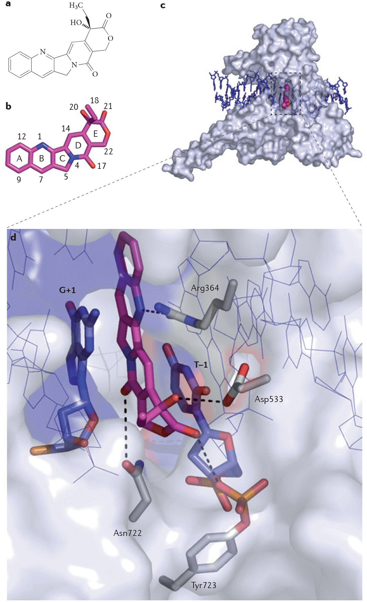
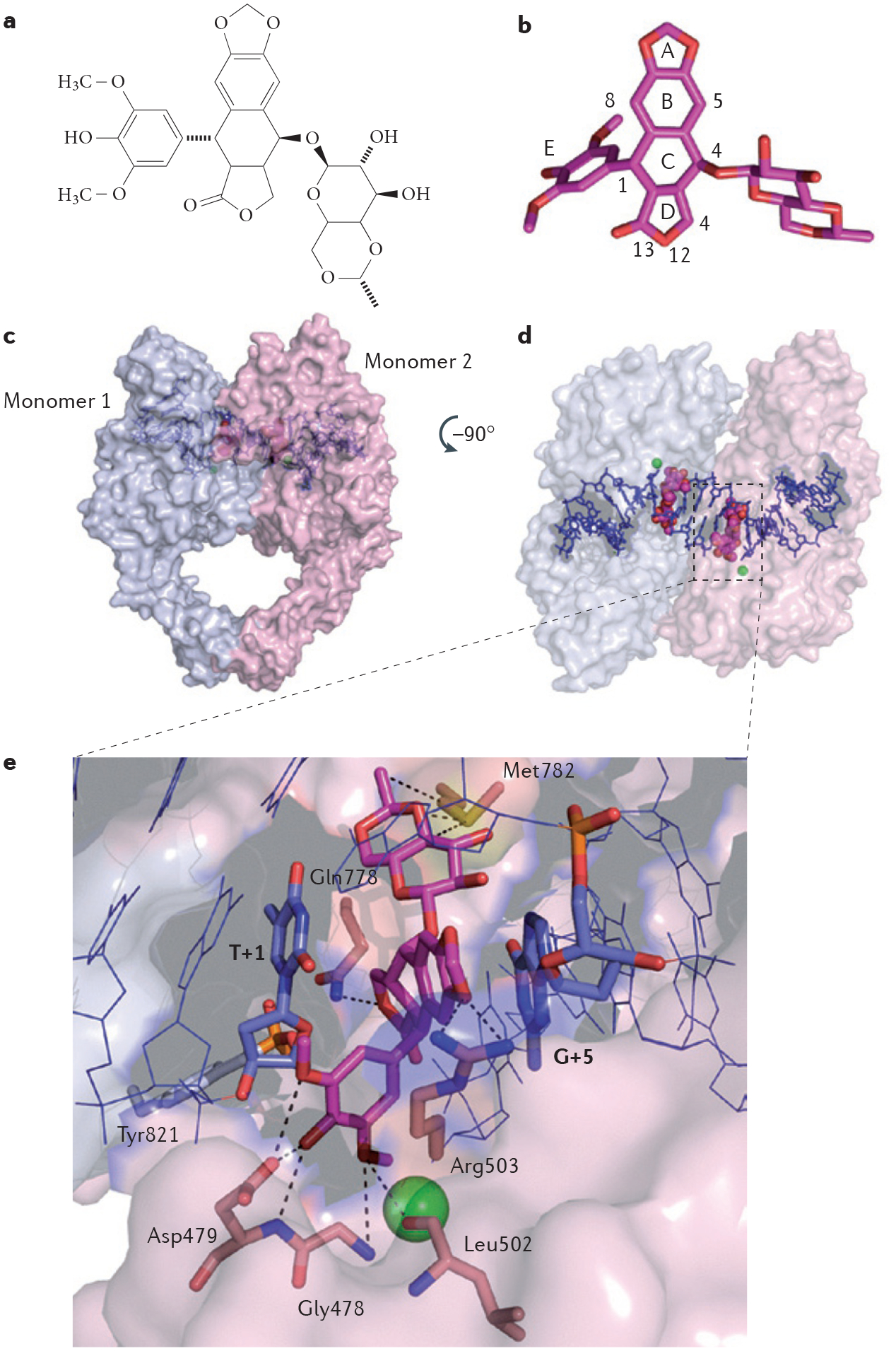
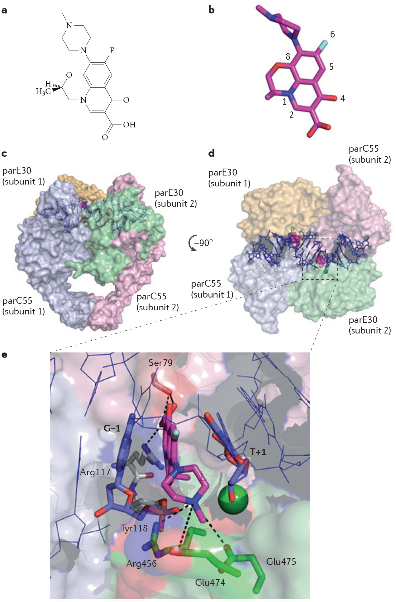
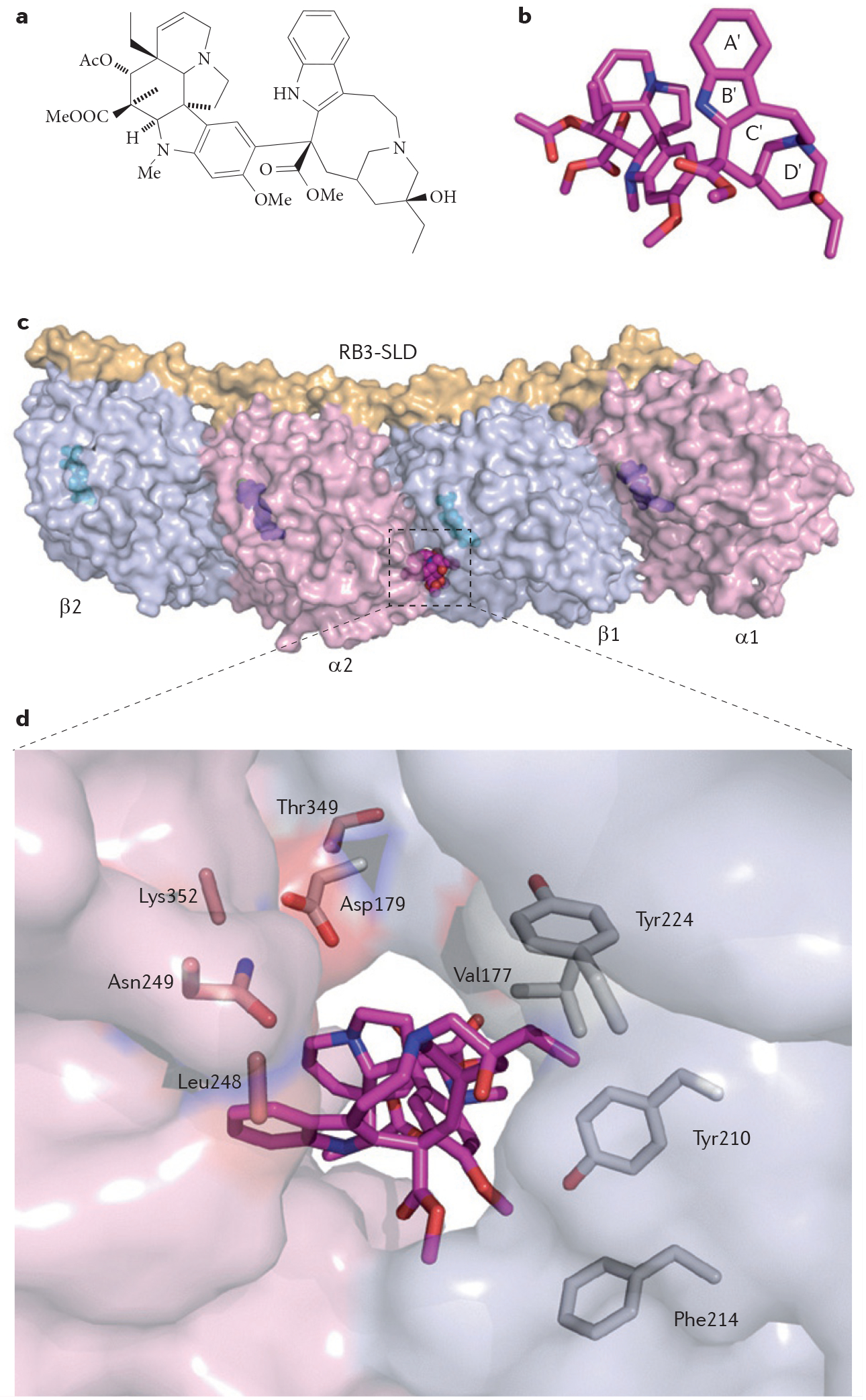
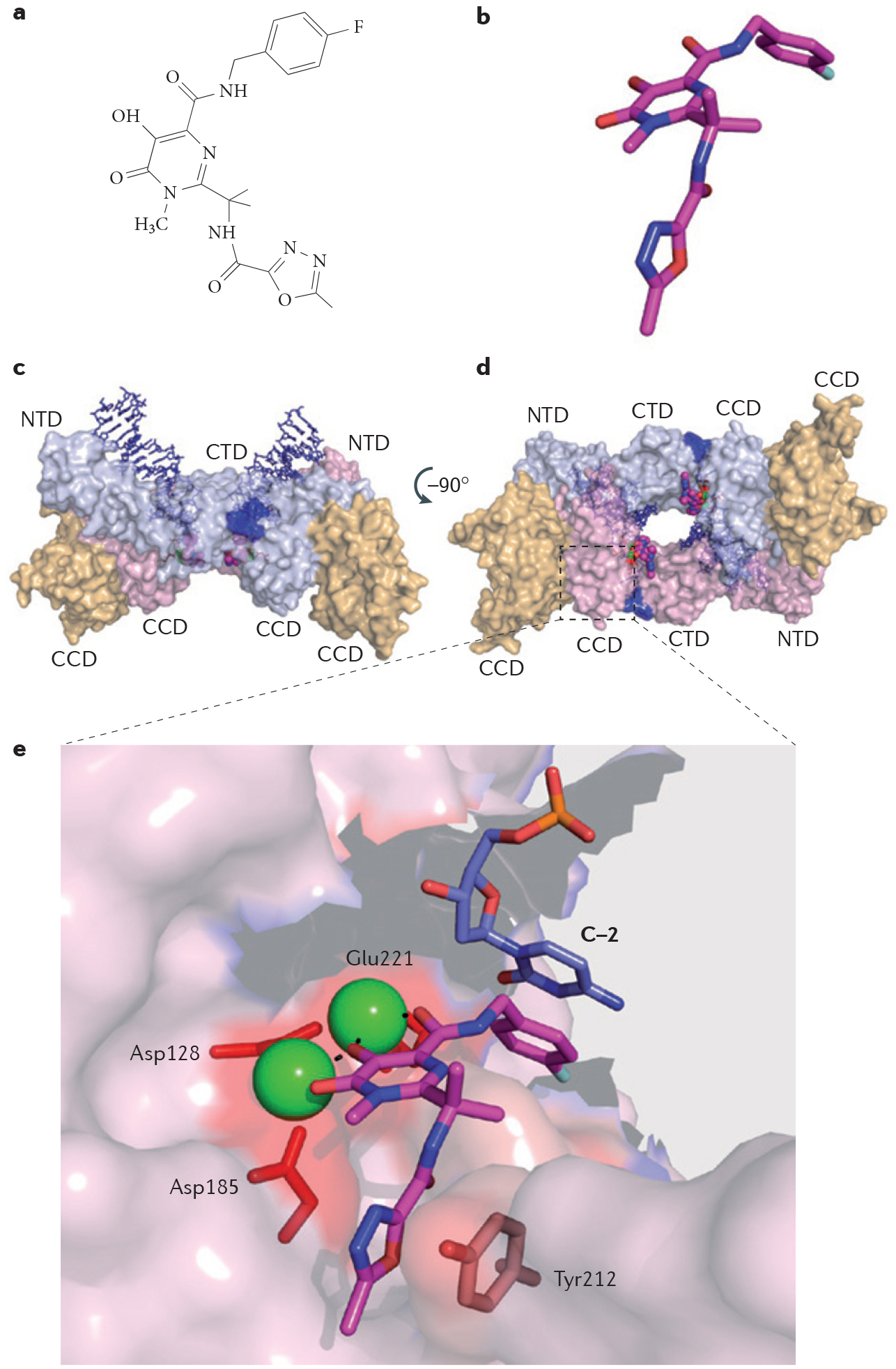
Interfacial inhibitors belong to a broad class of natural products and synthetic drugs that are commonly used to treat cancers as well as bacterial and HIV infections. They bind selectively to interfaces as macromolecular machines assemble and are set in motion. The bound drugs transiently arrest the targeted molecular machines, which can initiate allosteric effects, or desynchronize macromolecular machines that normally function in concert. Here, we review five archetypical examples of interfacial inhibitors: the camptothecins, etoposide, the quinolone antibiotics, the vinca alkaloids and the novel anti-HIV inhibitor raltegravir. We discuss the common and diverging elements between interfacial and allosteric inhibitors and give a perspective for the rationale and methods used to discover novel interfacial inhibitors.


Conflict of interest statement

Competing interests statement

The authors declare no competing financial interests.

Figures

Figure 1 |
Figure 1 |. Structure of a topoisomerase I cleavage complex trapped by camptothecin.
a | Chemical structure of camptothecin (CPT). b | Three-dimensional structure of CPT (stick representation with carbon, nitrogen and oxygen atoms coloured in magenta, blue and red, respectively; Protein Data Bank ID code 1T8I; numbering according to REF. 26). c | Topoisomerase I (TOP1) is shown in surface representation (grey) to highlight the depth of the CPT binding pocket. CPT (shown in magenta) is bound inside the catalytic core of TOP1, intercalated between the DNA base pairs (shown in blue) flanking the TOP1 cleavage site. This mechanism of action is common to other TOP1 poisons. d | Interaction network between CPT, DNA and TOP1 in the drug–TOP1–DNA ternary complex. DNA contacts by π-stacking with the flanking DNA base pairs (G+1 and T–1) are indicated in blue. Protein contacts are illustrated, with the following hydrogen bonds demonstrated using dashed lines: hydrogen bonding between the N1 nitrogen atom of CPT and the guanidinium group of Arg364; hydrogen bonding between the 20 hydroxyl group (see panel a) of CPT and the carboxylic functional group of Asp533; and hydrogen bonding between the C17 pyridone ring oxygen and the side chain nitrogen of Asn722.
Figure 2 |
Figure 2 |. Structure of a topoisomerase Iiβ cleavage complex trapped by etoposide.
a | Chemical structure of etoposide. b | Three-dimensional structure of etoposide (stick representation with carbon and oxygen coloured in magenta and red, respectively; Protein Data Bank ID code 3QX3 (REF. 16); numbering according to REF. 16). c | Topoisomerase IIβ (TOP2β) is shown in surface representation and functions as a homodimer (represented by the light blue and light pink subunits). Etoposide (shown in magenta) is bound inside the catalytic core of each TOP2β subunit and stabilizes the cleavage complex by intercalating between the DNA base pairs (shown in blue) flanking the TOP2β cleavage sites. d | The image shown is the surface representation of TOP2β (in the left panel) after 90° rotation. e | Interaction network between etoposide in the drug–TOP2β–DNA ternary complex. DNA contacts by π-stacking with the flanking DNA base pairs (T+1 on the cleaved strand and G+5 on the uncleaved strand) are indicated in blue. The active site of TOP2β is assembled in trans with the catalytic Tyr821 residue (shown in grey) from monomer 1 and the magnesium (green sphere)-chelating triad of acidic residues (not shown) from monomer 2. Protein contacts from monomer 2 with etoposide are shown, with the following hydrogen bonds and Van der Waals interactions depicted using dashed lines: hydrogen bonding and Van der Waals interactions between the E ring oxygen atoms of etoposide and Gly478, Asp479 and Leu502 residues; between the A ring oxygen of etoposide and the Arg503 residue; between oxygen 12 on the D ring of etoposide and the Gln778 residue; and between the glycosidic group of etoposide and the Met782 residue.
Figure 3 |
Figure 3 |. Structure of a topoisomerase IV cleavage complex trapped by the fluoroquinolone antibiotic levofloxacin.
a | Chemical structure of levofloxacin. b | Three-dimensional structure of levofloxacin (stick representation with carbon, nitrogen, oxygen and fluorine atoms coloured in magenta, blue, red and grey, respectively; Protein Data Bank ID code 3K9F,; numbering according to REF. 11). c | Bacterial topoisomerase IV functions as a tetramer of two parC55 (light blue and light pink) and two parE30 (light green and yellow) subunits, shown in surface representation. Levofloxacin (shown in magenta) stabilizes the cleavage complex by intercalating between the DNA base pairs (shown in blue) flanking the topoisomerase IV cleavage sites. d | The image shown is the surface representation of topoisomerase IV (in the left panel) after 90 rotation. e | Interaction network between levofloxacin in the ternary complex formed by the drug, topoisomerase IV and DNA. DNA contacts by π-stacking with the flanking DNA base pairs (G–1 and T+1) are indicated in blue. The active site of topoisomerase IV is assembled in trans with the catalytic Tyr118 residue (shown in grey) from the parC55 subunit 1 and the magnesium (green sphere)-chelating triad of acidic residues (not shown) from the parE30 subunit 2. Protein contacts are shown, with the following hydrogen bonds depicted using dashed lines: hydrogen bonding between the C3 carboxyl group of levofloxacin and the residues Ser79 and Arg117 of the parC55 subunit; and the hydrogen bonds involving the Arg456, Glu474 and Glu475 residues of the parE30 subunit, which hold together the N4 nitrogen atom of the piperazine ring at the other side of the levofloxacin molecule.
Figure 4 |
Figure 4 |. Structure of vinblastine bound in a ternary complex with tubulin heterodimers.
a | Chemical structure of vinblastine. b | Three-dimensional structure of vinblastine (in sphere representation with carbon, nitrogen and oxygen atoms coloured in magenta, blue and red, respectively; Protein Data Bank (PDB) ID code 1Z2B). Heterocycles labelled A′ to D′ correspond to the catharanthine domain, and the other portion of the molecule corresponds to the vindoline portion of vinblastine. c | Vinblastine binds between an α-subunit (α2; shown in light pink) and a β-subunit (β1; shown in light blue) of two tubulin α-β heterodimers, in complex with the Rhodopirellula baltica protein 3 (RB3) stathmin-like domain (SLD), as shown in surface representation. GDP and GTP molecules are shown in sphere representation in cyan and magenta, respectively. d | Interaction network between vinblastine and tubulin heterodimers. Vinblastine is buried inside the complex and its orientation allows its catharanthine and vindoline moieties to interact with both tubulin heterodimers. Following vinblastine binding, the β-subunit residue Asp179 and the α-subunit residue Thr349 from each heterodimer move towards the drug and contribute to its binding. Vinblastine is stabilized on one side by the α-subunit residues Leu248, Asn249 and Lys352. On the other side, it is stabilized by the β-subunit residues Val177, Tyr210, Phe214 and Tyr224.
Figure 5 |
Figure 5 |. Structure of the retroviral prototype foamy virus intasome in ternary complex with the strand transfer integrase inhibitor raltegravir.
a | Chemical structure of raltegravir. b | Stick representation of raltegravir with carbon, nitrogen and oxygen atoms coloured in magenta, blue and red, respectively; Protein Data Bank (PDB) ID code 3OYA,. c | Surface representation of the retroviral intasome formed by a dimer of prototype foamy virus integrase, two viral DNA ends (shown in blue) and two molecules of raltegravir (shown in magenta). Each full-length monomer (shown in light blue and light pink) consists of three domains: the core catalytic domain (CCD), the carboxy-terminal domain (CTD) and the amino-terminal domain (NTD), and is associated in the crystal structure with a structural CCD (shown in yellow). d | Surface representation of the intasome rotated 90 around the horizontal axis to reveal the site of integration into the host DNA, occupied by two molecules of raltegravir. e | Interaction network between raltegravir, viral DNA (stick representation; shown in blue) and integrase residues (shown in red). Raltegravir forms three types of contacts: chelation of the catalytic Mg2+ ions (represented by green spheres) via its carbonyl groups (the three catalytic acidic residues, Asp128, Asp185 and Glu221, that chelate the Mg2+ ion on the other side are represented by red sticks); DNA binding via π-π interactions with the penultimate base of the cleaved viral DNA (C–2; shown in blue); and integrase binding by π-π interactions with the Tyr212 residue.

References

    1. Conti C et al. Replication fork velocities at adjacent replication origins are coordinately modified during DNA replication in human cells. Mol. Biol. Cell 18, 3059–3067 (2007). - PMC - PubMed
    1. Sugino A, Peebles CL, Kreuzer KN & Cozzarelli NR Mechanism of action of nalidixic acid: purification of Escherichia coli nalA gene product and its relationship to DNA gyrase and a novel nicking-closing enzyme. Proc. Natl Acad. Sci. USA 74, 4767–4771 (1977). - PMC - PubMed
    1. Gellert M, Mizuuchi K, O’Dea MH, Itoh T & Tomizawa JI Nalidixic acid resistance: a second genetic character involved in DNA gyrase activity. Proc. Natl Acad. Sci. USA 74, 4772–4776 (1977). - PMC - PubMed
    1. Pommier Y, Leo E, Zhang H & Marchand C DNA topoisomerases and their poisoning by anticancer and antibacterial drugs. Chem. Biol 17, 421–433 (2010). - PMC - PubMed
    1. Staker BL et al. The mechanism of topoisomerase I poisoning by a camptothecin analog. Proc. Natl Acad. Sci. USA 99, 15387–15392 (2002). - PMC - PubMed

Publication types

MeSH terms

Substances