

Experimental in vivo acute and chronic biomechanical and histomorphometrical comparison of self-drilling and self-tapping anterior cervical screws
- PMID: 22173611
- PMCID: PMC3337909
- DOI: 10.1007/s00586-011-2120-z
Experimental in vivo acute and chronic biomechanical and histomorphometrical comparison of self-drilling and self-tapping anterior cervical screws
Abstract
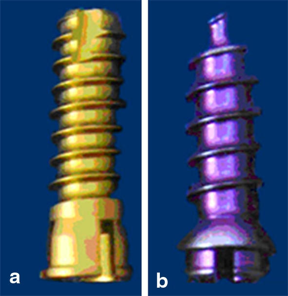
Purpose: Self-drilling screws (SDS) and self-tapping screws (STS) allow for quicker bone insertion and are associated with increased anchorage. This is an experimental in vivo comparison of anterior cervical SDS and STS in the post-insertion acute and chronic phases.
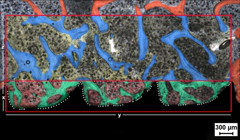
Methods: Thirty C2-C6 vertebrae from six Santa Inês hair sheep were used. Each screw design was randomly assigned to five of each spinal level. Insertion torque was measured using a torque device. Three animals were killed in each phase. Vertebrae were randomly assigned to pullout tests and histomorphometrical bone-screw interface evaluation (percent screw-bone contact and bone density inside and outside the threaded area). Statistical significance was set at P < 0.05.
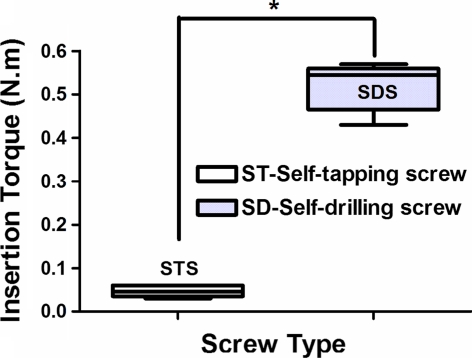
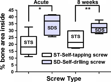
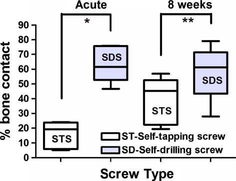
Results: SDS insertion torque was greater than STS (P = 0.0001). SDS pullout strength was significantly greater than STS in the acute and chronic phases (P = 0.0001, 0.0003, respectively). SDS percent screw-bone contact and inside area bone density were significantly greater in both phases. No outside area bone density differences were observed in either phase.
Conclusions: SDS had higher insertion torque and better anchorage than STS in both phases. SDS percent bone-screw contact and inside area bone density were higher in both phases.
Figures


Similar articles
-
Effect of pilot hole on biomechanical and in vivo pedicle screw-bone interface.Eur Spine J. 2013 Aug;22(8):1829-36. doi: 10.1007/s00586-013-2810-9. Epub 2013 May 8. Eur Spine J. 2013. PMID: 23653133 Free PMC article.
-
Factors affecting the pullout strength of self-drilling and self-tapping anterior cervical screws.Spine (Phila Pa 1976). 2003 Jan 1;28(1):9-13. doi: 10.1097/00007632-200301010-00004. Spine (Phila Pa 1976). 2003. PMID: 12544947
-
BIOMECHANICAL EVALUATION OF THE INFLUENCE OF CERVICAL SCREWS TAPPING AND DESIGN.Rev Bras Ortop. 2015 Dec 8;44(5):415-9. doi: 10.1016/S2255-4971(15)30272-X. eCollection 2009 Jan. Rev Bras Ortop. 2015. PMID: 27004189 Free PMC article.
-
Biomechanical analysis of bone mineral density, insertion technique, screw torque, and holding strength of anterior cervical plate screws.J Neurosurg. 1995 Aug;83(2):325-9. J Neurosurg. 1995. PMID: 7616279 Clinical Trial.
-
Biomechanics of cervical spine internal fixation.Spine (Phila Pa 1976). 1991 Mar;16(3 Suppl):S10-6. doi: 10.1097/00007632-199103001-00004. Spine (Phila Pa 1976). 1991. PMID: 2028323 Review.
Cited by
-
Using self-drilling screws in volar plate osteosynthesis for distal radius fractures: a feasibility study.BMC Musculoskelet Disord. 2016 Mar 10;17:120. doi: 10.1186/s12891-016-0972-4. BMC Musculoskelet Disord. 2016. PMID: 26966085 Free PMC article.
-
[Biomechanical changes of sheep cervical spine after unilateral hemilaminectomy and different degrees of facetectomy].Beijing Da Xue Xue Bao Yi Xue Ban. 2019 Aug 18;51(4):728-732. doi: 10.19723/j.issn.1671-167X.2019.04.023. Beijing Da Xue Xue Bao Yi Xue Ban. 2019. PMID: 31420630 Free PMC article. Chinese.
-
Effect of pilot hole on biomechanical and in vivo pedicle screw-bone interface.Eur Spine J. 2013 Aug;22(8):1829-36. doi: 10.1007/s00586-013-2810-9. Epub 2013 May 8. Eur Spine J. 2013. PMID: 23653133 Free PMC article.
References
Publication types
MeSH terms
LinkOut - more resources
Full Text Sources
Medical
Miscellaneous
