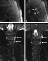
The role of MR myelography with intrathecal gadolinium in localization of spinal CSF leaks in patients with spontaneous intracranial hypotension
- PMID: 22173753
- PMCID: PMC7966438
- DOI: 10.3174/ajnr.A2815
The role of MR myelography with intrathecal gadolinium in localization of spinal CSF leaks in patients with spontaneous intracranial hypotension
Abstract
Background and purpose: Localization of spinal CSF leaks in CSF hypovolemia is critical in directing focal therapy. In this retrospective review, our aim was to determine whether GdM was helpful in confirming and localizing spinal CSF leaks in patients in whom no leak was identified on a prior CTM.
Materials and methods: Forty-one symptomatic patients with clinical suspicion of SIH were referred for GdM after undergoing at least 1 CTM between February 2002 and August 2010. A retrospective review of the imaging and electronic medical records was performed on each patient.
Results: In 17 of the 41 patients (41%), GdM was performed for follow-up of a previously documented leak at CTM. In the remaining 24 patients (59%), in whom GdM was performed for a suspected CSF leak, which was not identified on CTM, GdM localized the CSF leak in 5 of 24 patients (21%). In 1 of these 5 patients, GdM detected the site of leak despite negative findings on brain MR imaging, spine MR imaging, and CTM of the entire spine. Sixteen of 17 patients with previously identified leaks underwent interval treatment, and leaks were again identified in 12 of 17 (71%).
Conclusions: GdM is a useful technique in the highly select group of patients who have debilitating symptoms of SIH, a high clinical index of suspicion of spinal CSF leak, and no demonstrated leak on conventional CTM. Intrathecal injection of gadolinium contrast remains an off-label use and should be reserved for those patients who fail conventional CTM.
Figures


References
-
- Mokri B. Spontaneous cerebrospinal fluid leaks: from intracranial hypotension to cerebrospinal fluid hypovolemia—evolution of a concept. Mayo Clin Proc 1999; 74: 1113– 23 - PubMed
-
- Schievink WI. Spontaneous spinal cerebrospinal fluid leaks and intracranial hypotension. JAMA 2006; 295: 2286– 96 - PubMed
-
- Aydin K, Guven K, Sencer S, et al. MRI cisternography with gadolinium-containing contrast medium: its role, advantages and limitations in the investigation of rhinorrhoea. Neuroradiology 2004; 46: 75– 80. Epub 2003 Nov 13 - PubMed
-
- Aydin K, Terzibasioglu E, Sencer S, et al. Localization of cerebrospinal fluid leaks by gadolinium-enhanced magnetic resonance cisternography: a 5-year single-center experience. Neurosurgery 2008; 62: 584– 89, discussion 584–89 - PubMed
MeSH terms
Substances
LinkOut - more resources
Full Text Sources
Other Literature Sources
Medical
