

Investigation of unmedicated early onset restless legs syndrome by voxel-based morphometry, T2 relaxometry, and functional MR imaging during the night-time hours
- PMID: 22173758
- PMCID: PMC8050438
- DOI: 10.3174/ajnr.A2829
Investigation of unmedicated early onset restless legs syndrome by voxel-based morphometry, T2 relaxometry, and functional MR imaging during the night-time hours
Abstract
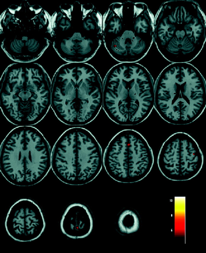
Background and purpose: The pathophysiology of eRLS has not yet been elucidated. The purpose of the study was to assess, in patients with eRLS, the volume, iron content, and activation of the brain during night-time episodes of SLD and PLMs.
Materials and methods: Eleven right-handed unmedicated patients with eRLS (mean age, 55.3 ± 8.4 years; disease duration, 17.5 ± 14.05 years) and 11 matched control subjects were studied with a T1-weighted high-resolution 3D spoiled gradient-echo sequence used for VBM and a multisection spin-echo T2-weighted sequence used for T2 relaxometry. Additionally, a single-shot multisection gradient echo-planar sequence was used for fMRI. Brain activation was recorded during spontaneous SLD and PLMs. SPM software was used for analysis of the functional data.
Results: The patients showed no regional brain volume change, but T2 relaxometry revealed decreased T2 relaxation time in the right globus pallidus internal and the STN, indicating increased iron content. The patients were observed to activate the following areas: in the left hemisphere, the primary motor and somatosensory cortex, the thalamus, the pars opercularis, and the ventral anterior cingulum; and in the right hemisphere, the striatum, the inferior and superior parietal lobules, and the dorsolateral prefrontal cortex. Bilateral activation was observed in the cerebellum, the midbrain, and the pons.
Conclusions: eRLS is associated with increased iron content of the globus pallidus internal and STN, suggesting dysfunction of the basal ganglia. Activation of the striatofrontolimbic area may represent the neurofunctional substrate mediating the repetitive compulsive movements seen in RLS.
Figures
References
-
- Garcia-Borreguero D. Time to REST: epidemiology and burden. Eur J Neurol 2006;13: 15–20 - PubMed
-
- Paulus W, Dowling P, Rijman R, et al. . Update of the pathophysiology of the restless-legs-syndrome. Mov Disord 2007;22: S431–39 - PubMed
-
- Connor JR, Boyer PJ, Menzies SL, et al. . Neuropathological examination suggests impaired brain iron acquisition in restless legs syndrome. Neurology 2003;61: 304–09 - PubMed
-
- Earley CJ, B Barker P, Horská A, et al. . MRI-determined regional brain iron concentrations in early- and late-onset restless legs syndrome. Sleep Med 2006;7: 458–61. Epub 2006 Jun 5 - PubMed
MeSH terms
Substances
LinkOut - more resources
Full Text Sources
Medical
Miscellaneous