

Evaluation of aqueductal stenosis by 3D sampling perfection with application-optimized contrasts using different flip angle evolutions sequence: preliminary results with 3T MR imaging
- PMID: 22173764
- PMCID: PMC8050433
- DOI: 10.3174/ajnr.A2833
Evaluation of aqueductal stenosis by 3D sampling perfection with application-optimized contrasts using different flip angle evolutions sequence: preliminary results with 3T MR imaging
Abstract
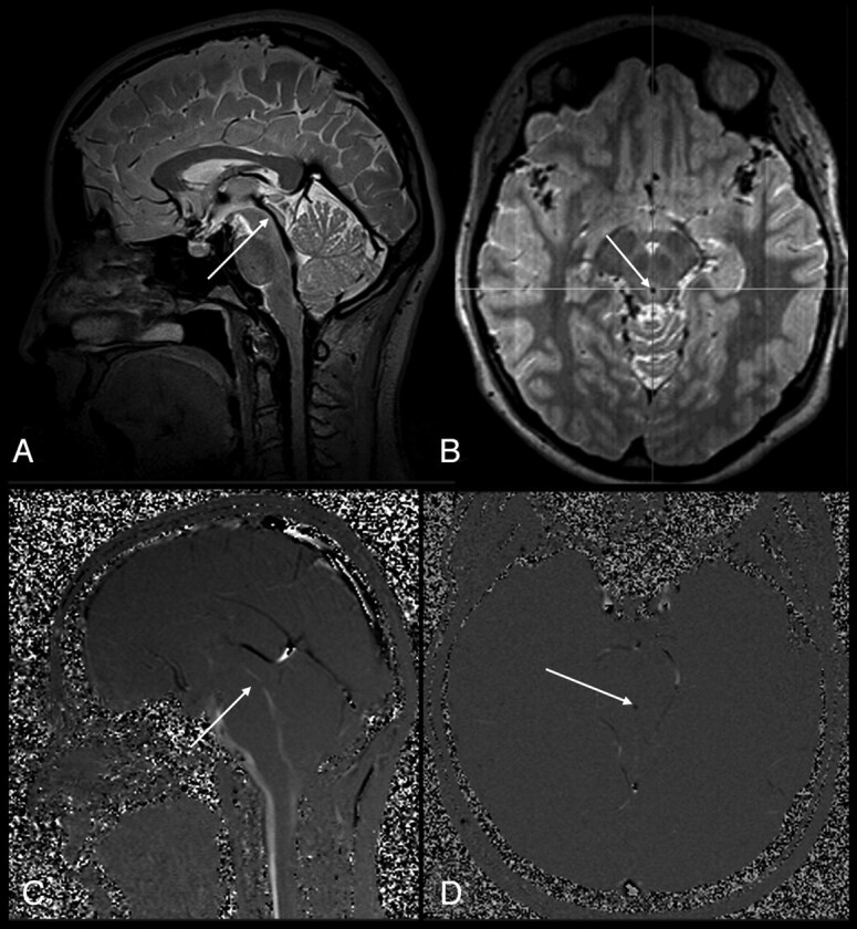
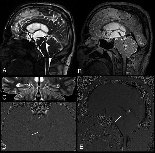
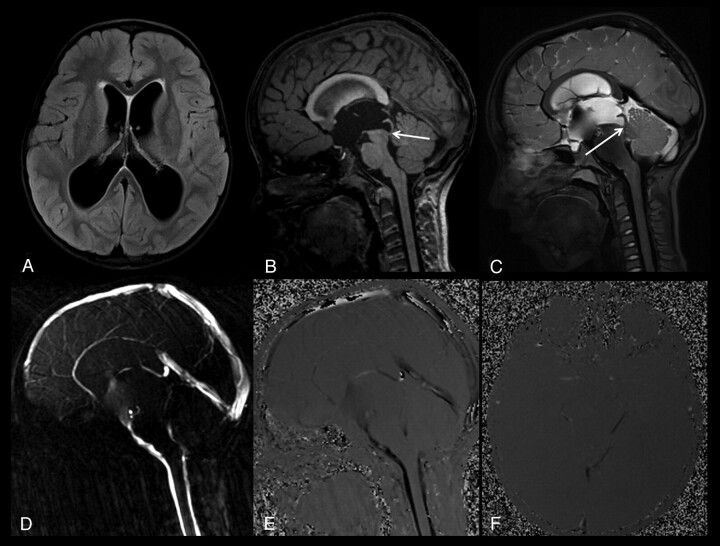
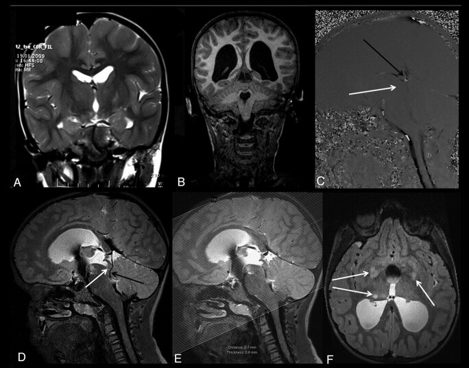
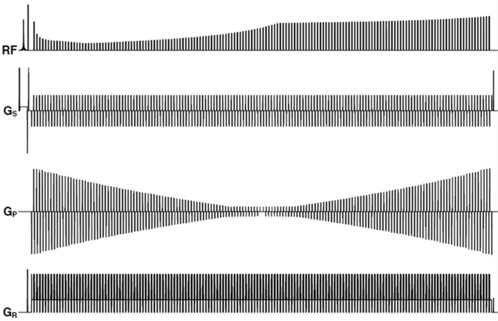
Background and purpose: Diagnosis of AS and periaqueductal abnormalities by routine MR imaging sequences is challenging for neuroradiologists. The aim of our study was to evaluate the utility of the 3D-SPACE sequence with VFAM in patients with suspected AS.
Materials and methods: PC-MRI and 3D-SPACE images were obtained in 21 patients who had hydrocephalus on routine MR imaging scans and had clinical suspicion of AS, as well as in 12 control subjects. Aqueductal patency was visually scored (grade 0, normal; grade 1, partial obstruction; grade 2, complete stenosis) by 2 experienced radiologists on PC-MRI (plus routine T1-weighted and T2-weighted images) and 3D-SPACE images. Two separate scores were statistically compared with each other as well as with the consensus scores obtained from general agreement of both radiologists.
Results: There was an excellent correlation between 3D-SPACE and PC-MRI scores (κ = 0.828). The correlation between 3D-SPACE scorings and consensus-based scorings was higher compared with the correlation between PC-MRI and consensus-based scorings (r = 1, P < .001 and r = 0.966, P < .001, respectively).
Conclusions: 3D-SPACE sequence with VFAM alone can be used for adequate and successful evaluation of the aqueductal patency without the need for additional sequences and examinations. Noninvasive evaluation of the whole cranium is possible in a short time with high resolution by using 3D-SPACE.
Figures
References
-
- Allan R, Chaseling R, Graf N, et al. Aqueduct stenosis-benign? J Clin Neurosci 2005;12:178–82 - PubMed
-
- Tisell M. How should primary aqueductal stenosis in adults be treated? A review. Acta Neuro Scand 2005;111:143–53 - PubMed
-
- Algin O, Hakyemez B, Parlak M. Phase-contrast MRI and 3D-CISS versus contrast-enhanced MR cisternography on the evaluation of the aqueductal stenosis. Neuroradiology 2010;52:99–108 - PubMed
-
- Sekerci Z, Akalan N, Kilic C, et al. Primary non-neoplastic aqueductal stenosis associated with Von Recklinghausen's disease. Turk Neurosurg 1989;1:30–33
-
- Algin O. Role of complex hydrocephalus in unsuccessful endoscopic third ventriculostomy. Childs Nerv Syst 2010;26:3–4 - PubMed
MeSH terms
LinkOut - more resources
Full Text Sources
Medical