

Kimura disease: CT and MR imaging findings
- PMID: 22173767
- PMCID: PMC8050465
- DOI: 10.3174/ajnr.A2854
Kimura disease: CT and MR imaging findings
Abstract
Background and purpose: KD is a rare chronic inflammatory disorder of unknown etiology. The purpose of this study was to evaluate the CT and MR imaging findings of KD in the head and neck.
Materials and methods: We retrospectively reviewed the CT (n = 21) and MR (n = 9) images obtained in 28 patients (24 males and 4 females; mean age, 32 years; age range, 10-62 years) with histologically proved KD in the head and neck.
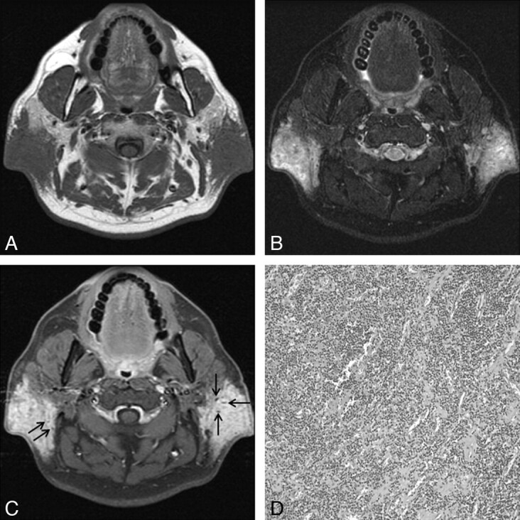
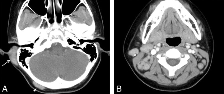
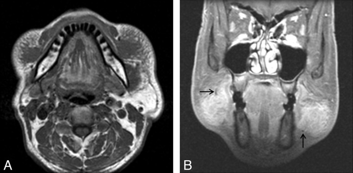
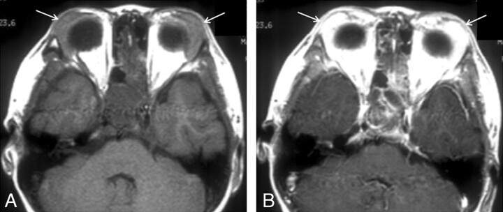
Results: In these 28 patients, CT and MR images demonstrated a total of 52 non-nodal lesions, 1-8 cm in greatest diameter, in the head and neck. The lesions were unilateral in 11 patients and bilateral in 17 patients. Eleven patients had a solitary lesion, and 17 patients had 2-4 lesions. The parotid and/or periparotid area was the most frequent location, with 36 lesions in 23 patients. The margin of the lesions was well-defined in 1 and ill-defined in 51 cases. Compared with the adjacent muscle, the MR signal intensity of all lesions was iso- to slightly hyperintense on T1-weighted images and hyperintense on T2-weighted images. Most of the lesions demonstrated mild or moderate enhancement on postcontrast CT scans and moderate or marked enhancement on postcontrast MR images. MR images also showed tubular signal-intensity voids in 7 of 13 lesions. Associated lymphadenopathy was demonstrated in 23 patients, usually bilaterally.
Conclusions: Multiple ill-defined enhancing masses within and around the parotid gland with associated regional lymphadenopathy are characteristic CT and MR imaging findings of KD in the head and neck.
Figures
Similar articles
-
Nodular fasciitis in the head and neck: CT and MR imaging findings.AJNR Am J Neuroradiol. 2005 Nov-Dec;26(10):2617-23. AJNR Am J Neuroradiol. 2005. PMID: 16286411 Free PMC article.
-
Castleman disease of the neck: CT and MR imaging findings.Eur J Radiol. 2014 Nov;83(11):2041-50. doi: 10.1016/j.ejrad.2014.08.013. Epub 2014 Sep 2. Eur J Radiol. 2014. PMID: 25223886
-
Kimura disease involving parotid gland and cervical nodes: CT and MR findings.J Comput Assist Tomogr. 1992 Mar-Apr;16(2):320-2. doi: 10.1097/00004728-199203000-00028. J Comput Assist Tomogr. 1992. PMID: 1545036
-
Head and neck lesions. Radiologic-pathologic correlations.Radiol Clin North Am. 1998 Sep;36(5):983-1014, vii. doi: 10.1016/s0033-8389(05)70072-5. Radiol Clin North Am. 1998. PMID: 9747197 Review.
-
Hypointense head and neck lesions on T2-weighted images: correlation with histopathologic findings.Neuroradiology. 2020 Oct;62(10):1207-1217. doi: 10.1007/s00234-020-02483-z. Epub 2020 Jun 20. Neuroradiology. 2020. PMID: 32562036 Review.
Cited by
-
Kimura disease forming a human polyomavirus 6-negative parotid gland nodule with prominent squamous metaplasia in a young female: A case report.Radiol Case Rep. 2023 Mar 16;18(5):1933-1938. doi: 10.1016/j.radcr.2023.02.027. eCollection 2023 May. Radiol Case Rep. 2023. PMID: 36970233 Free PMC article.
-
A Stepwise decision tree model for differential diagnosis of Kimura's disease in the head and neck.BMC Med Imaging. 2025 Mar 17;25(1):90. doi: 10.1186/s12880-025-01618-z. BMC Med Imaging. 2025. PMID: 40097945 Free PMC article.
-
Kimura disease in children: A report of 11 cases and review of the literature.Front Pediatr. 2023 Feb 17;11:1131963. doi: 10.3389/fped.2023.1131963. eCollection 2023. Front Pediatr. 2023. PMID: 36873634 Free PMC article.
-
Clinical analysis of Kimura's disease in 24 cases from China.BMC Surg. 2020 Jan 2;20(1):1. doi: 10.1186/s12893-019-0673-7. BMC Surg. 2020. PMID: 31898499 Free PMC article.
-
Oropharyngeal Kimura's disease: a diagnostic dilemma and therapeutic challenge.BMJ Case Rep. 2020 Oct 30;13(10):e236366. doi: 10.1136/bcr-2020-236366. BMJ Case Rep. 2020. PMID: 33127696 Free PMC article.
References
-
- Kuo TT, Shih LY, Chan HL.. Kimura's disease: involvement of regional lymph nodes and distinction from angiolymphoid hyperplasia with eosinophilia. Am J Surg Pathol 1988;12: 843–54 - PubMed
-
- Chen H, Thomson LD, Aguilera NS, et al. . Kimura disease: a clinopathologic study of 21 cases. Am J Surg Pathol 2004;28: 505–13 - PubMed
-
- Kimura T, Yoshimura S, Ishikawa E.. Unusual granulation combined with hyperplastic changes of lymphatic tissue [in Japanese]. Trans Soc Pathol Jpn 1948;37: 179–80
-
- Kung IT, Gibson JB, Bannatyne PM.. Kimura's disease: a clinicopathological study of 21 cases and its distinction from angiolymphoid hyperplasia with eosinophilia. Pathology 1984;16: 39–44 - PubMed
-
- Li TJ, Chen XM, Wang S-Z, et al. . Kimura's disease: a clinicopathologic study of 54 Chinese patients. Oral Surg Oral Med Oral Pathol Oral Radiol Endod 1996;82: 549–55 - PubMed
Publication types
MeSH terms
LinkOut - more resources
Full Text Sources
Medical