. 2012 May;113(5):1599-607.
doi: 10.1002/jcb.24028.

Effect of phorbol 12-myristate 13-acetate activated signaling pathways on 1α, 25 dihydroxyvitamin D3 regulated human 25-hydroxyvitamin D3 24-hydroxylase gene expression in differentiated Caco-2 cells

Affiliations

Effect of phorbol 12-myristate 13-acetate activated signaling pathways on 1α, 25 dihydroxyvitamin D3 regulated human 25-hydroxyvitamin D3 24-hydroxylase gene expression in differentiated Caco-2 cells

Yan Jiang et al. J Cell Biochem. 2012 May.

Abstract

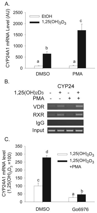
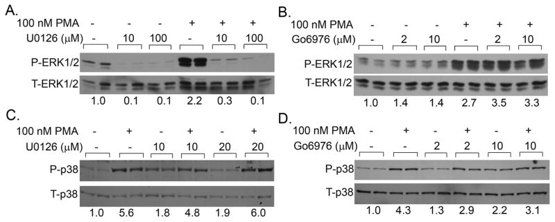
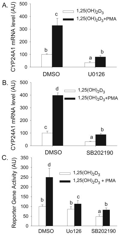
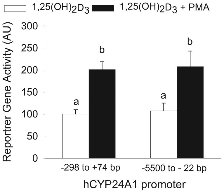
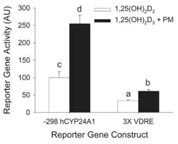
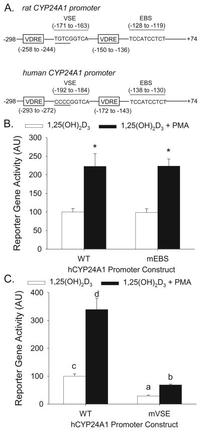
Phorbol-12-myristate-13-acetate (PMA), a protein kinase C(PKC) activator, can modulate 1α, 25 dihydroxyvitamin D(3) (1,25(OH)(2)D(3))-induced expression of the 24-hydroxylase (CYP24A1) gene but this has not been studied in differentiated enterocytes, a primary 1,25(OH)(2) D(3) target cell. We found that in differentiated Caco-2 cells, an established model of the mature absorptive epithelial cell, PMA significantly enhanced 1,25(OH)(2)D(3)-induced human CYP24A1 (hCYP24A1) mRNA accumulation and hCYP24A1 promoter-luciferase reporter gene activation by 150%. Reporter gene studies further identified the region between -298 and +74 bp in the hCYP24A1 promoter as critical for the PMA enhancing effect and chromatin immunoprecipitation (ChIP) analysis showed that PMA enhanced 1,25(OH)(2)D(3)-induced binding of vitamin D receptor to this region. PMA can activate PKC, ERK1/2, and p38 MAP kinases and inhibition of these signaling pathways reduced both 1,25(OH)(2)D(3)-induced hCYP24A1 gene transcription and the enhancing effect of PMA. The PMA enhancing effect on 1,25(OH)(2)D(3) action was evident in a minimal promoter with three osteocalcin VDREs and was reduced after mutation of a putative vitamin D stimulatory site in the hCYP24A1 promoter. In contrast, mutation of a Ets binding site (EBS) in the hCYP24A1 promoter had no impact on 1,25(OH)(2)D(3) action or the PMA enhancing effect. These data suggest that in the differentiated enterocyte PMA-induced activation of several signaling pathways contribute to 1,25(OH)(2)D(3)-induced hCYP24A1 gene expression through multiple regulatory motifs within the proximal hCYP24A1 promoter.


Figures

Figure 1
Figure 1
(A) Effect of PMA on 1,25(OH)2D3-induced CYP24A1 mRNA expression in differentiated Caco-2 cells. Cells were pretreated for 5 min with 100 nM PMA followed by treatment for 2 h with 10 nM 1,25(OH)2D3 or vehicle in the absence of PMA. Data are expressed as mean±sem (n=4) with the vehicle treated group as a reference value (100). (B) Effect of PMA on 1,25(OH)2D3-induced association of VDR and RXR on the proximal human CYP24A1 gene promoter in differentiated Caco-2 cells. ChIP assays were performed with non-immune rabbit immunoglobulin (IgG), or antibodies against VDR or RXR. ChIP enriched DNA was amplified with primers recognizing the proximal CYP24 promoter (−252 to −52 bp). (C) Effect of PKC inhibition on PMA enhanced CYP24A1 induction by 1,25(OH)2D3. Cells were pretreated with 10 μM Go6976 for 30 min and 100 nM PMA was added for the last 5 min. Afterwards inhibitor and PMA were removed and cells were treated with 10 nM 1, 25(OH)2D3 or vehicle in the absence of PMA. Data are expressed relative to the 1,25(OH)2D3-induced expression of CYP24A1 (calculated as 1,25(OH)2D3/vehicle = 100). Bars with different letter superscripts are significantly different from one another (p< 0.05, Fisher’s protected LSD). Data are representative of three independent experiments.
Figure 2
Figure 2
Effect of PMA on MAPK activation in differentiated Caco-2 cells. (A,B) Effect of MEK (U0126) or PKC inhibition (Go6976) on PMA-induced ERK1/2 phosphorylation. Cells were treated with inhibitors or vehicle for 30 min and PMA or vehicle was added for the last 5 min. Proteins from whole cell extracts were isolated and analyzed for total and phosphorylated ERK1/2. (C,D) Effect of MEK (U0126) or PKC inhibition (Go6976) on PMA-induced p38 kinase phosphorylation. Cells were pretreated with inhibitors or vehicle for 30 min and PMA or vehicle was added for the last 5 min. Inhibitor treatments were continued for an additional 1h in the absence of PMA. Proteins from whole cell extracts were isolated and analyzed for total and phosphorylated p38 kinase. Data are representative of three independent experiments. Number on the bottom is the ratio between phosphorylated and total protein levels.
Figure 3
Figure 3
Inhibitors of ERK1/2, classic PKC and p38 kinase independently reduce 1,25(OH)2D3-induced and PMA-enhanced CYP24A1 gene expression in differentiated Caco-2 cells. (A, B) Cells were preincubated with inhibitors (A: 10 μM U0126, B: 8 μM SB202190) for 30 min and PMA (100 nM) or vehicle was added for the last 5 min. Afterwards inhibitor treatment was continued with 10 nM 1,25(OH)2D3 or ethanol vehicle for an additional 2 h in the absence of PMA. (C) Cells were transfected with a −298 to +74 bp hCYP24A1 promoter-luciferase reporter gene. 15 h after transfection, cells were pre-treated with or without inhibitors (10 μM U0126,8 μM SB202190) for 30 min and PMA (100 nM) or vehicle was added for the last 10 min. Afterwards inhibitor treatment was continued with 10 nM 1,25(OH)2D3 or ethanol vehicle for an additional 4 h in the absence of PMA. Data are expressed as mean±sem (n=4) relative to the 1,25(OH)2D3-induced expression of CYP24A1 (calculated as 1,25(OH)2D3/vehicle = 100). Bars with different letter superscripts are significantly different from one another (P< 0.05, Fisher’s protected LSD). Data in each panel are representative of three independent experiments.
Figure 4
Figure 4
The proximal CYP24A1 promoter contains the elements necessary for enhancement of 1,25(OH)2D3 transactivation by PMA. Differentiated Caco-2 cells were transfected with one of two different hCYP24A1 promoter-luciferase plasmids (−298 to +74 bp or −5500 to −22 bp). 15 h after transfection, cells were pre-treated with 100 nM PMA for 10 min followed by 10 nM 1,25(OH)2D3 for 4 h in the absence of PMA. Data are expressed as mean±sem (n=6) relative to the 1,25(OH)2D3-induced expression of the −298 to +74 bp hCYP24A1 promoter (calculated as 1,25(OH)2D3/vehicle = 100). Bars with different letter superscripts are significantly different from one another (P< 0.05, Fisher’s protected LSD).
Figure 5
Figure 5
The enhancement of PMA is partially mediated through VDREs in the CYP24A1 promoter. Differentiated Caco-2 cells were transfected with a −298 to +74 bp- human CYP24A1 promoter-luciferase or 3X osteopontin VDRE thymidine kinase-luciferase (3X VDRE) reporter gene construct. 15 h after transfection cells were pre-treated with 100 nM PMA for 10 min followed by vehicle or 10 nM 1, 25(OH)2D3 for 4 h in the absence of PMA. Data are expressed as mean±sem (n=4) relative to the 1,25(OH)2D3-induced expression of the −298 hCYP24A1 promoter (calculated as 1,25(OH)2D3/vehicle = 100). Bars with different letter superscripts are significantly different from one another (P<0.05, Fisher’s protected LSD). Data are representative of three independent experiments.
Figure 6
Figure 6
A VSE site but not the EBS site is involved in 1,25(OH)2D3-induced and PMA-enhanced human CYP24A1 promoter activity in differentiated Caco-2 cells. (A) Schematics showing the location of the EBS (Ets protein binding site) and VSE (vitamin D stimulation element) in the rat and human CYP24A1 promoters. Differentiated Caco-2 cells were transfected with wild type (WT), EBS mutated (B, mEBS) or VSE mutated (C, mVSE) −298 to +74 bp human CYP24A1 promoter-luciferase reporter gene constructs. 15 h after transfection, cells were pre-treated with 100 nM PMA for 10 mins followed by 10 nM 1, 25(OH)2D3 for 4 h in the absence of PMA. Data are expressed as mean±sem (n=6) relative to the 1,25(OH)2D3-induced expression of the wild-type −298 to 74 bp hCYP24A1 promoter (calculated as 1,25(OH)2D3/vehicle = 100). In (A) * = Significantly different from the 1,25(OH)2D3-induced group within each reporter gene construct (p< 0.05, Fisher’s protected LSD). In (B) Bars with different letter superscripts are significantly different from one another (P<0.05, Fisher’s protected LSD). Data are representative of three independent experiments.

Similar articles

Cited by

References

    1. Almog T, Naor Z. Mitogen activated protein kinases (MAPKs) as regulators of spermatogenesis and spermatozoa functions. Molecular and Cellular Endocrinology. 2008;282:39–44. - PubMed
    1. Armbrecht HJ, Boltz MA, Hodam TL, Kumar VB. Differential responsiveness of intestinal epithelial cells to 1,25- dihydroxyvitamin D3--role of protein kinase C. J Endocrinol. 2001;169:145–151. - PubMed
    1. Armbrecht HJ, Chen ML, Hodam TL, Boltz MA. Induction of 24-hydroxylase cytochrome P450 by 1,25-dihydroxyvitamin D and phorbol exters in normal rat kidney (NRK-52E) cells. J Endocrinol. 1997;153:199–205. - PubMed
    1. Armbrecht HJ, Hodam TL, Boltz MA, Chen ML. Phorbol ester markedly increases the sensitivity of intestinal epithelial cells to 1,25-dihydroxyvitamin D3. FEBS Lett. 1993;327:13–16. - PubMed
    1. Barletta F, Dhawan P, Christakos S. Integration of hormone signaling in the regulation of human 25(OH)D3 24-hydroxylase transcription. Am J Physiol Endocrinol Metab. 2004;286:E598–E608. - PubMed

Publication types

MeSH terms

LinkOut - more resources