Review
. 2011 Dec;189(4):1145-75.
doi: 10.1534/genetics.111.128264.

Regulation of cell wall biogenesis in Saccharomyces cerevisiae: the cell wall integrity signaling pathway

Affiliations
Review

Regulation of cell wall biogenesis in Saccharomyces cerevisiae: the cell wall integrity signaling pathway

David E Levin. Genetics. 2011 Dec.

Abstract

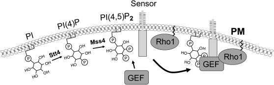
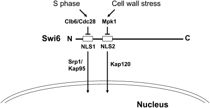
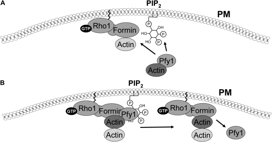
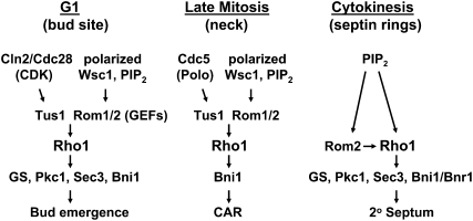
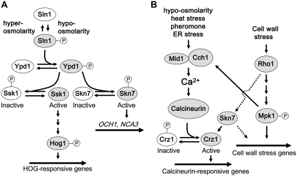
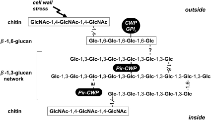
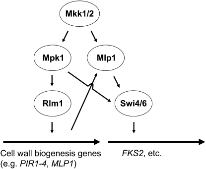
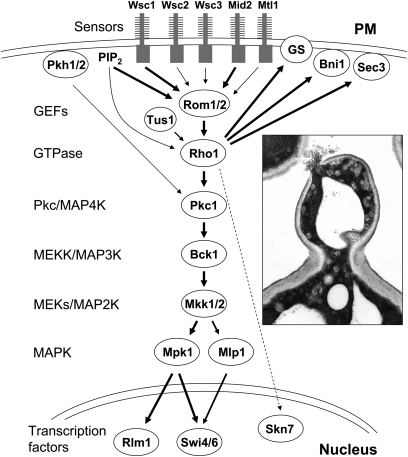
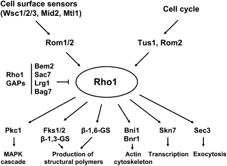
The yeast cell wall is a strong, but elastic, structure that is essential not only for the maintenance of cell shape and integrity, but also for progression through the cell cycle. During growth and morphogenesis, and in response to environmental challenges, the cell wall is remodeled in a highly regulated and polarized manner, a process that is principally under the control of the cell wall integrity (CWI) signaling pathway. This pathway transmits wall stress signals from the cell surface to the Rho1 GTPase, which mobilizes a physiologic response through a variety of effectors. Activation of CWI signaling regulates the production of various carbohydrate polymers of the cell wall, as well as their polarized delivery to the site of cell wall remodeling. This review article centers on CWI signaling in Saccharomyces cerevisiae through the cell cycle and in response to cell wall stress. The interface of this signaling pathway with other pathways that contribute to the maintenance of cell wall integrity is also discussed.


Figures

Figure 1
Figure 1
Molecular organization of the yeast cell wall (adapted from Lesage and Bussey 2006, doi: 10.1128/MMBR.00038-05; amended with permission from American Society for Microbiology). Chains of β-1,3-glucan, branched through β-1,6-linkages, form a mesh network that provides the mechanical strength of the cell wall and also serves as a scaffold for the attachment of cell wall proteins (CWPs). Pir-CWPs are attached directly to β-1,3-glucan through a Gln residue within their internal repeats that is converted to a Glu (E) residue in the linkage. These proteins have the potential to cross-link β-1,3-glucan chains through multiple repeat sequences. GPI-CWPs are attached to the network indirectly through a linkage between the lipidless GPI remnant (GPIr) and β-1,6-glucan. Chitin, a polymer of β-1,4-N-actetylglucosamine (GlcNAc), can be attached either directly to β-1,3-glucan on the inner surface or indirectly by β-1,6-glucan to the outer surface. The latter attachment is induced in response to cell wall stress. The nature of the linkage between β-1,3-glucan and β-1,6-glucan chains is still uncharacterized.
Figure 2
Figure 2
The CWI signaling pathway. Signals are initiated at the plasma membrane (PM) through the cell-surface sensors Wsc1, -2, -3, Mid2, and Mtl1. The extracellular domains of these proteins are highly O-mannosylated. Together with PIP2, which recruits the Rom1/2 GEFs to the plasma membrane, the sensors stimulate nucleotide exchange on Rho1. Relative input of each sensor is indicated by the width of the arrows. Additional regulatory inputs from the Tus1 GEF and the Pkh1/2 protein kinases are indicated. The various effectors of Rho1 include the β-1,3-glucan synthase (GS), β-1,6-glucan synthase activity (not shown), formins (Bni1), Sec3, and the Pkc1-activated MAPK cascade. Mlp1 is a pseudokinase paralog of Mpk1 that contributes to the transcriptional program through a noncatalytic mechanism. Two transcription factors, Rlm1 and SBF (Swi4/Swi6), are activated by the pathway. Skn7 (dashed line) may also contribute to the CWI transcriptional program. (Inset) Thin-section electron micrograph of a Pkc1-depleted cell that has undergone cell lysis at its bud tip. Conditions were as described in Levin et al. (1994).
Figure 3
Figure 3
Rho1 regulators and effectors. Rho1 localization and activity are regulated through the cell cycle and in response to cell wall stress by cell-surface sensors, a family of GEFs (Rom1, Rom2, and Tus1), and a set of GAPs (Bem2, Sac7, Lrg1, and Bag7). Six known Rho1 effectors control cell wall biogenesis through polymer synthesis, polarization of the actin cytoskeleton, directed secretion, and transcription.
Figure 4
Figure 4
The phosphoinositide-signaling pathway at the plasma membrane (PM). The sequential action of Stt4 and Mss4 at the cell surface generates PI(4,5)P2 (PIP2), which recruits Rho1-GEFs to the PM through their PH domains for interaction with the cytoplasmic tails of the cell-surface sensors.
Figure 5
Figure 5
The involvement of PIP2 in the delivery of actin to the Rho1–formin complex. (A) Profilin is an actin-binding protein that delivers actin to the actin-nucleating formins Bni1 and Bnr1. The profilin–actin complex is recruited to the PM by PIP2, where it is bound by the active Rho1–formin complex. At least one additional Rho1 effector, Sec3, is also recruited to the PM by PIP2 (not shown). (B) Upon delivery of actin to the formin, PIP2 is thought to stimulate the release of actin from profilin, thereby driving actin polymerization.
Figure 6
Figure 6
Control of the Skn7 transcription factor. (A) The Sln1 branch of the HOG pathway. The Sln1 osmosensor controls a phosphorelay pathway, which activates Skn7 under hypo-osmotic conditions to support cell wall biosynthesis and the Hog1 MAPK cascade under hyper-osmotic conditions. Active components are shaded. (B) Coordinate activation of CWI signaling, Ca2+ signaling, and Skn7. Rho1 may independently activate the Skn7 transcription factor (dashed line), which induces stabilization of the Crz1 transcripion factor, and may have additional effects on cell wall stress-induced transcription. The Mid1-Cch1 Ca2+ channel is activated by many of the same stresses that activate CWI signaling. Additionally, activation of Mpk1 results in stimulation of the Mid1-Cch1 Ca2+ channel, at least in response to ER stress, which activates the Ca2+-dependent protein phosphatase calcineurin. The Crz1 transcription factor is activated through dephosphorylation by calcineurin, which allows its entry to the nucleus. This interplay may coordinate control of gene expression by Ca2+ signaling and cell wall stress signaling.
Figure 7
Figure 7
The CWI pathway transcriptional program. The majority of genes regulated by CWI signaling are under the control of the Rlm1 transcription factor, which is phosphorylated and activated by Mpk1. Among these genes is MLP1, which encodes a pseudokinase paralog of Mpk1. Using a mechanism that is independent of protein kinase catalytic activity, Mpk1, together with Mlp1, drive expression of a subset of cell wall stress-induced genes through the Swi4/Swi6 transcription factor (including FKS2).
Figure 8
Figure 8
Model for Mpk1-driven FKS2 transcription. (A) Under non-inducing conditions, weak transcription initiation is attenuated by association of the Sen1–Nrd1–Nab3 termination complex to the elongating RNA Pol II. (B) Under inducing conditions, Mpk1 and Mlp1 (not shown) are activated in response to phosphorylation by Mkk1/2. (C) The active MAPK or pseudokinase binds to Swi4. (D) These dimers are competent to bind the FKS2 promoter. (E) Swi6 is recruited to form an Mpk1–Swi4–Swi6 complex on the FKS2 promoter. (F) RNA Pol II and the Paf1C are recruited to the promoter in a Swi6-dependent manner, completing formation of the initiation complex. (G) Mpk1 associates with Paf1, likely by hand off from Swi4. (H) Mpk1 overcomes transcriptional attenuation by blocking recruitment of the termination complex. SCB, Swi4-binding site.
Figure 9
Figure 9
Control of Swi6 nucleocytoplasmic shuttling. Swi6 possesses two nuclear localization (NLS) signals, NLS1 and NLS2, which are both regulated by phosphorylation. NLS1 is regulated through the cell cycle, and its function is blocked by phosphorylation on Ser160 by the S-phase CDK, Clb6/Cdc28, resulting in cytoplasmic localization of Swi6 at times other than G1. NLS2 is regulated by cell wall stress and its function is blocked by Mpk1 phosphorylation on Ser238. This feedback inhibitory event down-regulates cell wall stress-induced transcription after activation. Together, these two disparate signals converge to control Swi6 nuclear localization under different conditions. The indicated karyopherins recognize each NLS.
Figure 10
Figure 10
Control of Rho1 activity through the cell cycle. Rho1 is activated at three sites through the cell cycle: the incipient bud site and bud tip during wall expansion, the mother/bud neck during mitosis, and between the septin rings during cytokinesis. Rho1’s recruitment and activation at these sites involves different regulators and is likely to result in the activation of only a subset of effectors. The secondary septum is cell wall material that is distinct from the primary septum, which is chitin produced by Chs2. GS, glucan synthase; CAR, contractile actin ring.

References

    1. Abe J., Kusuhara M., Ulevitch R. J., Berk B. C., Lee J. D., 1996. Big mitogen-activated protein kinase 1 (BMK1) is a redox-sensitive kinase. J. Biol. Chem. 271: 16586–16590 - PubMed
    1. Abe M., Qadota H., Hirata A., Ohya Y., 2003. Lack of GTP-bound Rho1p in secretory vesicles of Saccharomyces cerevisiae. J. Cell Biol. 162: 85–97 - PMC - PubMed
    1. Adamo J. E., Rossi G., Brennwald P., 1999. The Rho GTPase Rho3 has a direct role in exocytosis that is distinct from its role in actin polarity. Mol. Biol. Cell 10: 4121–4133 - PMC - PubMed
    1. Aguilar-Uscanga B., François J. M., 2003. A study of the yeast cell wall composition and structure in response to growth conditions and mode of cultivation. Lett. Appl. Microbiol. 37: 268–274 - PubMed
    1. Alberts A. S., 2001. Identification of a carboxyl-terminal diaphanous-related formin homology protein autoregulatory domain. J. Biol. Chem. 276: 2824–2830 - PubMed

Publication types

MeSH terms