. 2012 Feb 10;287(7):4904-13.
doi: 10.1074/jbc.M111.307264. Epub 2011 Dec 15.

Phosphosite mapping of P-type plasma membrane H+-ATPase in homologous and heterologous environments

Affiliations

Phosphosite mapping of P-type plasma membrane H+-ATPase in homologous and heterologous environments

Elena L Rudashevskaya et al. J Biol Chem. .

Abstract

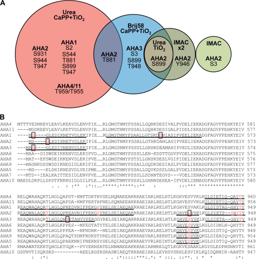
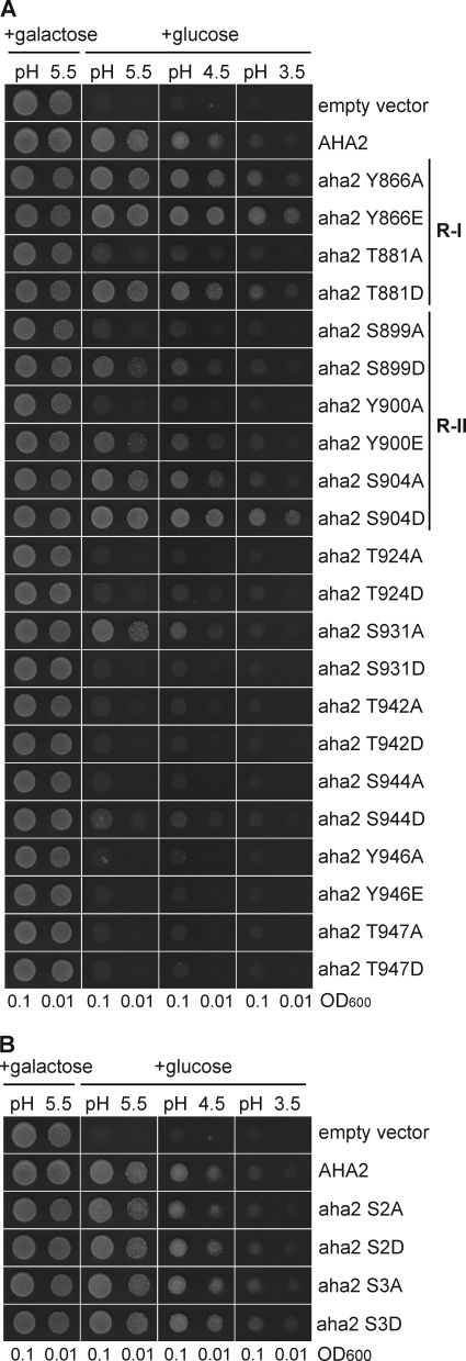
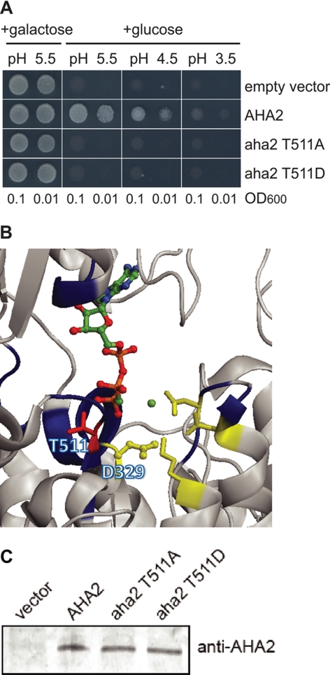
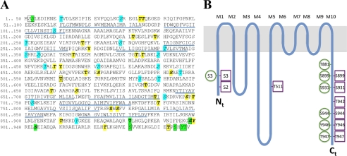
Phosphorylation is an important posttranslational modification of proteins in living cells and primarily serves regulatory purposes. Several methods were employed for isolating phosphopeptides from proteolytically digested plasma membranes of Arabidopsis thaliana. After a mass spectrometric analysis of the resulting peptides we could identify 10 different phosphorylation sites in plasma membrane H(+)-ATPases AHA1, AHA2, AHA3, and AHA4/11, five of which have not been reported before, bringing the total number of phosphosites up to 11, which is substantially higher than reported so far for any other P-type ATPase. Phosphosites were almost exclusively (9 of 10) in the terminal regulatory domains of the pumps. The AHA2 isoform was subsequently expressed in the yeast Saccharomyces cerevisiae. The plant protein was phosphorylated at multiple sites in yeast, and surprisingly, seven of nine of the phosphosites identified in AHA2 were identical in the plant and fungal systems even though none of the target sequences in AHA2 show homology to proteins of the fungal host. These findings suggest an unexpected accessibility of the terminal regulatory domain of plasma membrane H(+)-ATPase to protein kinase action.


Figures

FIGURE 1.
FIGURE 1.
Phosphosites identified in plasma membrane H+-ATPases homologously expressed in A thaliana membranes. A, phosphosites were determined in A. thaliana plasma membrane H+-ATPases using different approaches for phosphopeptide enrichment. Plasma membranes purified from A. thaliana seedlings were exposed for trypsin digestion directly, after treatment with Brij58, or after solubilization by urea/thiourea. IMAC, TiO2, and calcium phosphate precipitation followed by TiO2 (CaPP+TiO2) were used for phosphopeptide enrichment. Phosphorylation sites in the phosphopeptides were validated manually and are unique for the isoform of AHA mentioned in this figure with following exceptions: Site Ser-931 of AHA2 was determined in the peptide GHVEpSVVK, which can derive also from AHA2, AHA3, AHA5, AHA6, AHA8, AHA9, and Thr-959/955 in the peptide GLDIETIQQAYpTV that is shared by AHA4 and AHA11 isoforms. All the phosphopeptides determined are presented in the supplemental material. B, shown is ClustalW analysis of parts of the N terminus (N), P domain (P), and C terminus (C) of the ATPase isoforms of Arabidopsis. The identified phosphopeptides are underlined, and phosphorylated residues printed in bold and red. Novel residues are boxed.
FIGURE 2.
FIGURE 2.
Analysis of the importance of phosphosites in AHA2 for the activity of the pump by functional complementation of yeast pma1. The yeast strain RS-72 is dependent on the activity of the H+-ATPase expressed when grown on glucose media. When grown on galactose the endogenous yeast H+-ATPase PMA1 is expressed. A, shown is an analysis of pumps mutated in C-terminal phosphosites. B, shown is an analysis of pumps mutated in the N-terminal phosphosites.
FIGURE 3.
FIGURE 3.
Phosphorylation of recombinant autoinhibited plasma membrane H+-ATPase 2 (AHA2) heterologously expressed in yeast membranes. A, shown is a Euler diagram of the number of phosphosites determined by digesting of AHA2 in a mixture of solubilized membrane proteins with trypsin, Lys-C, and Glu-C. Trypsin and Lys-C were of the same effectiveness and gave one unique site each. B, shown is a summary table of phosphosites in AHA2 determined using complementary digestion by three enzymes, their distribution within the H+-ATPase domains, and number of cases (n) when phosphosites were determined within a total number of 7 samples analyzed. a/a, amino acid. Almost all the phosphosites determined are in the N- and C-terminal ends of the pump.
FIGURE 4.
FIGURE 4.
Phosphorylation of highly conservative Thr-551 situated in the catalytic center inhibits pump activity. A, shown is abolishment of yeast growth complementation when mutant in Thr-511 AHA2 was expressed. B, in the crystal structure of AHA2, Thr-511 is situated close to Asp-329, the conserved Asp phosphorylated during catalysis (59). C, shown is a Western blot analysis of expression level of the mutant AHA2 in yeast cells.
FIGURE 5.
FIGURE 5.
Overview of phosphosites in AHA2. A, shown is the amino acid sequence of AHA2. Green color, phospho-residues identified in AHA2 expressed homologously in planta (this study and previous studies). Boxed, phosphosites identified in recombinant AHA2 expressed heterologously in yeast. Italics (and cyan when not phosphorylated in AHA2 in planta), Phosphosites were predicted by the NetPhosK server. Bold (and yellow when not phosphorylated in AHA2 in planta), phosphosites predicted by the PhosPhAt 3.0 server; underlined, membrane spanning domains in the crystal structure of AHA2 (59). B, shown is a spaghetti model of AHA2 with the phosphosites indicated. Green circles, phosphosites when expressed in planta. Magenta squares, phosphosites when expressed in yeast.

References

    1. Cohen P. (2000) The regulation of protein function by multisite phosphorylation. A 25-year update. Trends Biochem. Sci. 25, 596–601 - PubMed
    1. Ptacek J., Snyder M. (2006) Charging it up. Global analysis of protein phosphorylation. Trends Genet. 22, 545–554 - PubMed
    1. Mann M., Jensen O. N. (2003) Proteomic analysis of post-translational modifications. Nat. Biotechnol. 21, 255–261 - PubMed
    1. Ozlu N., Akten B., Timm W., Haseley N., Steen H., Steen J. A. (2010) Phosphoproteomics. Wiley Interdiscip. Rev. Syst. Biol. Med. 2, 255–276 - PubMed
    1. Pawson T., Nash P. (2000) Protein-protein interactions define specificity in signal transduction. Genes Dev. 14, 1027–1047 - PubMed

Publication types

MeSH terms

Substances

LinkOut - more resources