. 2012 Jan 15;188(2):559-68.
doi: 10.4049/jimmunol.1100335. Epub 2011 Dec 14.

Helminth protection against autoimmune diabetes in nonobese diabetic mice is independent of a type 2 immune shift and requires TGF-β

Affiliations

Helminth protection against autoimmune diabetes in nonobese diabetic mice is independent of a type 2 immune shift and requires TGF-β

Marc P Hübner et al. J Immunol. .

Abstract

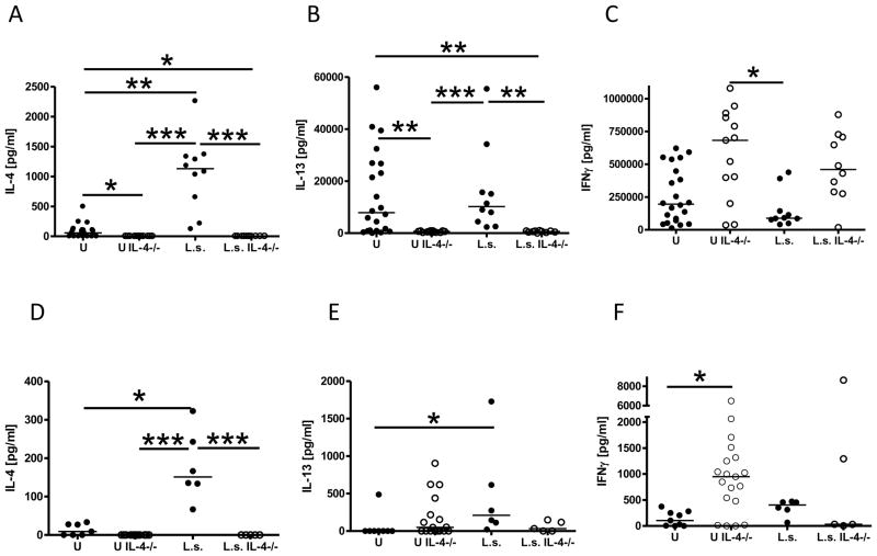
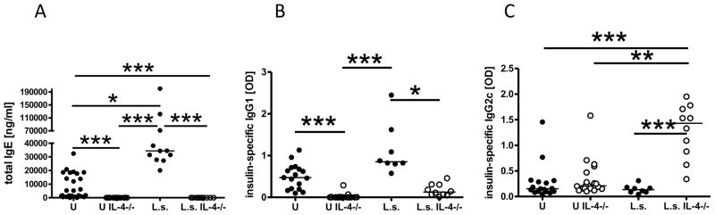
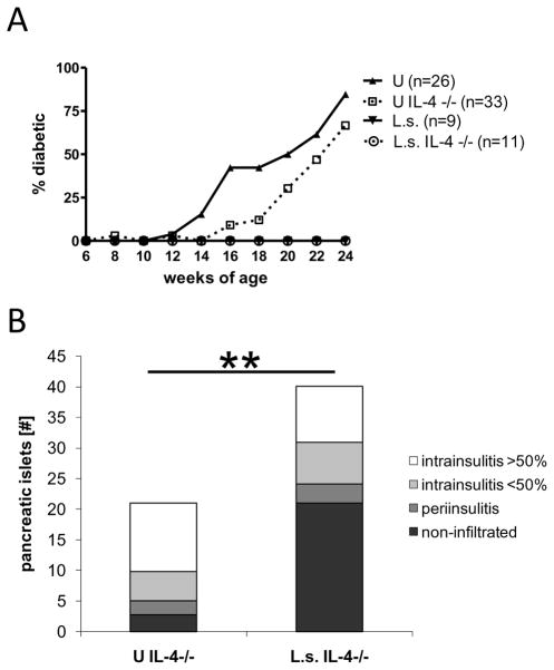
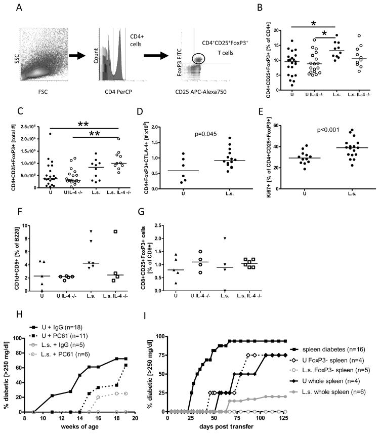
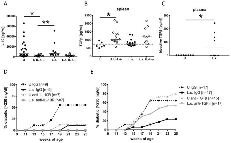
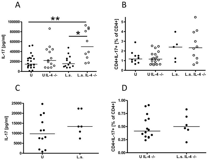
Leading hypotheses to explain helminth-mediated protection against autoimmunity postulate that type 2 or regulatory immune responses induced by helminth infections in the host limit pathogenic Th1-driven autoimmune responses. We tested these hypotheses by investigating whether infection with the filarial nematode Litomosoides sigmodontis prevents diabetes onset in IL-4-deficient NOD mice and whether depletion or absence of regulatory T cells, IL-10, or TGF-β alters helminth-mediated protection. In contrast to IL-4-competent NOD mice, IL-4-deficient NOD mice failed to develop a type 2 shift in either cytokine or Ab production during L. sigmodontis infection. Despite the absence of a type 2 immune shift, infection of IL-4-deficient NOD mice with L. sigmodontis prevented diabetes onset in all mice studied. Infections in immunocompetent and IL-4-deficient NOD mice were accompanied by increases in CD4(+)CD25(+)Foxp3(+) regulatory T cell frequencies and numbers, respectively, and helminth infection increased the proliferation of CD4(+)Foxp3(+) cells. However, depletion of CD25(+) cells in NOD mice or Foxp3(+) T cells from splenocytes transferred into NOD.scid mice did not decrease helminth-mediated protection against diabetes onset. Continuous depletion of the anti-inflammatory cytokine TGF-β, but not blockade of IL-10 signaling, prevented the beneficial effect of helminth infection on diabetes. Changes in Th17 responses did not seem to play an important role in helminth-mediated protection against autoimmunity, because helminth infection was not associated with a decreased Th17 immune response. This study demonstrates that L. sigmodontis-mediated protection against diabetes in NOD mice is not dependent on the induction of a type 2 immune shift but does require TGF-β.


Figures

FIGURE 1
FIGURE 1. IL-4-deficient NOD mice fail to develop type 2 cytokine responses during L. sigmodontis infection
Type 1 and type 2 cytokine production from splenocytes and pancreatic lymph node cells of uninfected (U) and L. sigmodontis-infected (L.s.) immunocompetent and IL-4-deficient NOD mice 1–2 weeks after the controls developed diabetes. Splenic production of (A) IL-4, (B) IL-13, (C) IFNγ, and production of (D) IL-4, (E) IL-13, (F) IFNγ from pancreatic lymph node cells that were stimulated with anti-CD3/anti-CD28. Each dot represents one mouse. Data is joined from 2–4 independent experiments. Statistical significance was assessed using the Kruskal-Wallis test, followed by Dunn’s post-hoc multiple comparisons. *p<0.05; **p<0.01; ***p<0.001
FIGURE 2
FIGURE 2. Helminth-infected IL-4 deficient NOD mice develop a predominant type 1 shift in antibody isotype profiles
A, plasma levels of total IgE (ng/ml) and (B) insulin-specific IgG1 as well as (C) insulin-specific IgG2c (OD) of uninfected (U) and L. sigmodontis-infected (L.s.) immunocompetent and IL-4-deficient NOD mice 1–2 weeks after the controls developed diabetes. Each dot represents one mouse. Data is joined from 2–4 independent experiments. Statistical significance was assessed using the Kruskal-Wallis test, followed by Dunn’s post-hoc multiple comparisons. *p<0.05; **p<0.01; ***p<0.001
FIGURE 3
FIGURE 3. Helminth-infection prevents the development of diabetes in NOD mice even in the absence of IL-4
A, percentages of diabetic mice (glucose >230 mg/dl) over time. Shown are immunocompetent and IL-4-deficient NOD mice infected with L. sigmodontis (L.s.) or corresponding uninfected controls (U). B, mean total numbers of pancreatic islets from IL-4-deficient L. sigmodontis infected NOD mice and uninfected controls (n=10 per group) 1–2 weeks after the controls developed diabetes. Pancreatic islets were classified as non-infiltrated, periinsulitis, and intrainsulitis with less than or greater than 50% infiltrated lymphocytes. Data is joined from 4 independent experiments. Significant differences between groups were analyzed by the Mann-Whitney test (** p<0.01).
FIGURE 4
FIGURE 4. Regulatory T cells are not required for L. sigmodontis-mediated protection against Type 1 diabetes
A, gating strategy for flow cytometric identification of regulatory T cells. Lymphocytes were gated by forward- (FSC) and sidescatter (SSC) characteristics (left panel). CD4 positive lymphocytes were gated (middle panel) and analyzed for CD25 and FoxP3 positivity (right panel). B, frequency and C, total number of splenic CD4+CD25+FoxP3+ T cells obtained from L. sigmodontis-infected (L.s.) and uninfected (U) IL-4-competent and IL-4-deficient NOD mice 1–2 weeks after the controls developed diabetes. D, total number of CD4+FoxP3+ spleen cells that express CTLA-4 in response to anti-CD3/anti-CD28. E, frequency of CD4+CD25+FoxP3+ spleen cells that spontaneously express the proliferation marker Ki67. F, frequency of splenic CD1dCD5+B220+ regulatory B cells and G, CD8+CD25+FoxP3+ regulatory T cells obtained from L. sigmodontis-infected (L.s.) and uninfected (U) IL-4-competent and IL-4-deficient NOD mice 1–2 weeks after the controls developed diabetes. H, Percentages of L. sigmodontis (L.s.) infected and uninfected (U) NOD mice that developed diabetes (glucose >250 mg/dl) while receiving anti-CD25 (PC61) or isotype control (IgG) weekly from 9 weeks of age to 20 weeks of age. I, Percentages of NOD.scid mice that developed diabetes after spleen cell transfer. NOD.scid mice received either splenocytes from diabetic NOD mice (squares), splenocytes from 14-week-old uninfected NOD mice (closed diamond), splenocytes depleted of FoxP3+ cells from 14-week-old uninfected NOD mice, splenocytes from L. sigmodontis-infected 14-week-old mice (open circles), or splenocytes depleted of FoxP3+ cells from 14-week-old L. sigmodontis-infected NOD mice. Each dot represents one mouse. Data for Fig. 4 A–E is joined from 2–4 independent experiments, F, G represents data from a single experiment, H and I, are the joined results from two and three independent experiments, respectively. Statistical significance was assessed using the Kruskal-Wallis test, followed by Dunn’s post-hoc multiple comparisons. Differences between two unpaired groups were tested for significance with the one-tailed Mann-Whitney test. *p<0.05; **p<0.01; ***p<0.001
FIGURE 5
FIGURE 5. Helminth-mediated protection against diabetes onset requires TGFβ
A, Production of IL-10 and (B) TGFβ in response to anti-CD3/anti-CD28 by splenocytes from uninfected (U) and L. sigmodontis-infected (L.s.) immunocompetent and IL-4-deficient NOD mice 1–2 weeks after the controls developed diabetes. The dashed line shows the baseline TGFβ concentration of the culture media. C, bioactive TGFβ levels in the plasma of L. sigmodontis-infected (L.s.) and uninfected NOD mice at 10 weeks of age. Development of diabetes (glucose >230 mg/dl) in mice that were either treated repeatedly with anti-IL-10R (D), anti-TGFβ (E), or isotype control. Shown are NOD mice infected with L. sigmodontis (L.s.) or corresponding uninfected controls (U). Each dot represents one mouse. Data for Fig. 5 A and B is joined from 2–4 independent experiments, C and D show results from one experiment, E shows joined results from two independent experiments. Statistical significance was assessed using the Kruskal-Wallis test, followed by Dunn’s post-hoc multiple comparisons. *p<0.05; **p<0.01
FIGURE 6
FIGURE 6. Helminth infection of NOD mice does not reduce the Th17 immune response
A, splenocyte production of IL-17 and (B) frequency of CD4+IL-17+ cells from spleens of uninfected (U) and L. sigmodontis-infected (L.s.) immunocompetent and IL-4-deficient NOD mice after activation with anti-CD3/anti-CD28. C, IL-17 release after anti-CD3/anti-CD28 stimulation of pancreatic lymph node cells of immunocompetent NOD mice. D, frequencies of CD4+CD17+ pancreatic lymph node cells from IL-4-deficient NOD mice. Each dot represents one mouse. Data is joined from 2–4 independent experiments. Statistical significance was assessed using the Kruskal-Wallis test, followed by Dunn’s post-hoc multiple comparisons. *p<0.05; **p<0.01

References

    1. Hübner MP, Stocker JT, Mitre E. Inhibition of type 1 diabetes in filaria-infected non-obese diabetic mice is associated with a T helper type 2 shift and induction of FoxP3+ regulatory T cells. Immunology. 2009;127:512–522. - PMC - PubMed
    1. Cooke A, Tonks P, Jones FM, O’Shea H, Hutchings P, Fulford AJ, Dunne DW. Infection with Schistosoma mansoni prevents insulin dependent diabetes mellitus in non-obese diabetic mice. Parasite Immunol. 1999;21:169–176. - PubMed
    1. Correale J, Farez M. Association between parasite infection and immune responses in multiple sclerosis. Ann Neurol. 2007;61:97–108. - PubMed
    1. Elliott DE, Setiawan T, Metwali A, Blum A, Urban JF, Jr, Weinstock JV. Heligmosomoides polygyrus inhibits established colitis in IL-10-deficient mice. Eur J Immunol. 2004;34:2690–2698. - PubMed
    1. Fleming JO, Cook TD. Multiple sclerosis and the hygiene hypothesis. Neurology. 2006;67:2085–2086. - PubMed

Publication types

MeSH terms