

Comparative evaluation of 18F-FDOPA, 13N-AMMONIA, 18F-FDG PET/CT and MRI in primary brain tumors - A pilot study
- PMID: 22174511
- PMCID: PMC3237222
- DOI: 10.4103/0972-3919.90256
Comparative evaluation of 18F-FDOPA, 13N-AMMONIA, 18F-FDG PET/CT and MRI in primary brain tumors - A pilot study
Abstract
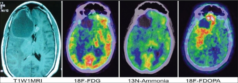
Aim: To determine the diagnostic reliability of 18F-FDOPA, 13N-Ammonia and 18F-FDG PET/CT in primary brain tumors and comparison with magnetic resonance imaging (MRI).
Materials and methods: A total of 23 patients, 8 preoperative and 15 postoperative, undergoing evaluation for primary brain tumors were included in this study. Of them, 9/15 were operated for high grade gliomas (7/9 astrocytomas and 2/9 oligodendrogliomas) and 6/15 for low grade gliomas (5/6 astrocytomas and 1/6 oligodendroglioma). After PET study, 2 of 8 preoperative cases were histopathologically proven to be of benign etiology. 3 low grade and 2 high grade postoperative cases were disease free on 6 months follow-up. Tracer uptake was quantified by standardized uptake values (SUV(max)) and the SUV max ratio of tumor to normal symmetrical area of contra lateral hemisphere (T/N). 18F-FDOPA uptake was also quantified by SUV(max) ratio of tumor to striatum (T/S). Conventional MR studies were done in all patients.
Results: Both high-grade and low-grade tumors were well visualized with 18F-FDOPA PET. Sensitivity of 18F-FDOPA PET was substantially higher (6/6 preoperative, 3/3 low grade postoperative, 7/7 high grade postoperative) than with 18F-FDG (3/6 preoperative, 1/3 low grade postoperative, 3/7 high grade postoperative) and 13N-Ammonia PET (2/6 preoperative, 1/3 low grade postoperative, 1/7 high grade postoperative). FDOPA was equally specific as FDG and Ammonia PET in operated cases but was falsely positive in two preoperative cases. Sensitivity of FDOPA (16/16) was more than MRI (13/16).
Conclusion: 18F-FDG uptake correlates with tumor grade. Though 18F-FDOPA PET cannot distinguish between tumor grade, it is more reliable than 18F-FDG and 13N-Ammonia PET for evaluating brain tumors. 18F-FDOPA PET may prove to be superior to MRI in evaluating recurrence and residual tumor tissue. 13N-Ammonia PET did not show any encouraging results.
Keywords: 13N-Ammonia; 18F-FDG; 18F-FDOPA; MRI; PET; brain tumors; sensitivity.
Conflict of interest statement
Figures

References
-
- Dooms GC, Hecht S, Brant-Zawadzki M, Berthiaume Y, Norman D, Newton TH. Brain radiation lesions: MR imaging. Radiology. 1986;15:149–55. - PubMed
-
- Van Dellen JR. Danziger A Failure of computerized tomography to differentiate between radiation necrosis and cerebral tumour. S Afr Med J. 1978;53:171–2. - PubMed
-
- Nelson SJ, Huhn S, Vigneron DB, Day MR, Wald LL, Prados M, et al. Volume MRI and MRSI techniques for the quantitation of treatment response in brain tumors: presentation of a detailed case study. J Magn Reson Imaging. 1997;7:1146–52. - PubMed
