

Crossed cerebellar diaschisis on F-18 FDG PET/CT
- PMID: 22174518
- PMCID: PMC3237210
- DOI: 10.4103/0972-3919.90263
Crossed cerebellar diaschisis on F-18 FDG PET/CT
Abstract
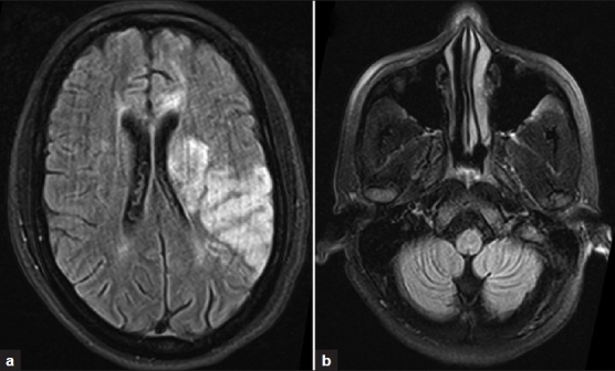
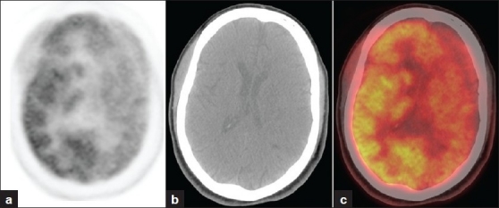
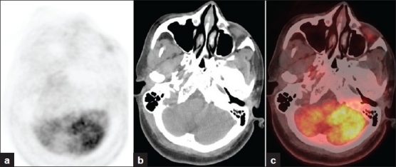
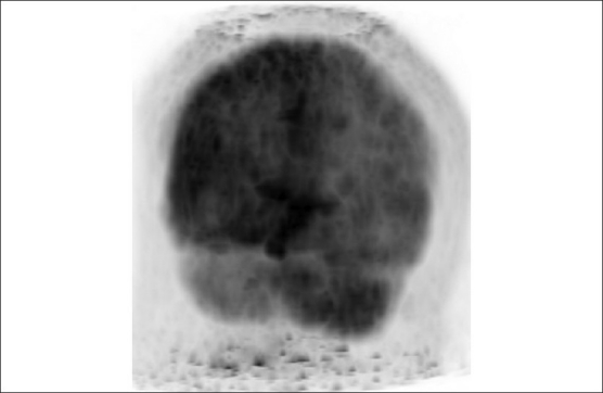
Diaschisis is the inhibition of function produced by focal disturbances in a portion of the brain at a distance from original site of injury. Many studies using brain SPECT (single-photon emission computed tomography) have demonstrated crossed cerebellar diaschisis (CCD) in patients with cerebral cortical infarct. We report a case of cerebrovascular accident involving the left middle cerebral artery territory. PET/CT performed one month after stroke showed hypometabolism in the left cerebral hemisphere with hypometabolism of the contralateral cerebellum. The finding of diminished glucose metabolism in the contralateral cerebellum represents CCD.
Keywords: Crossed cerebellar diaschisis; PET/CT; stroke.
Conflict of interest statement
Figures
References
-
- Baron JC, Bousser MG, Comar D, Castaigne P. Crossed cerebellar diaschisis in human supratentorial brain infarction. Trans Am Neurol Assoc. 1981;105:459–61. - PubMed
-
- Pantano P, Baron JC, Samson Y, Bousser MG, Derouesne C, Comar D. Crossed cerebellar diaschisis: Further studies. Brain. 1986;109:677–94. - PubMed
-
- Ito H, Kanno I, Shimosegawa E, Tamura H, Okane K, Hatazawa J. Hemodynamic changes during neural deactivation in human brain: A positron emission tomography study of crossed cerebellar diaschisis. Ann Nucl Med. 2002;16:249–54. - PubMed
-
- Wiesendanger M. Constantin von Monakow (1853–1930): A pioneer in interdisciplinary brain research and a humanist. Comptes Rendus Biologies. 2006;329:406–18. - PubMed
-
- Otte A, Roelcke U, von Ammon K, Hausmann O, Maguire RP, Missimer J, et al. Crossed cerebellar diaschisis and brain tumor biochemistry studied with positron emission tomography, [F18] fluorodeoxyglucose and [11 C] methionine. J Neurol Sci. 1998;156:73–7. - PubMed
