

Enucleated eyes after failed intra-arterial infusion of chemotherapy for unilateral retinoblastoma: histopathologic evaluation of vitreous seeding
- PMID: 22174572
- PMCID: PMC3236709
- DOI: 10.2147/OPTH.S24318
Enucleated eyes after failed intra-arterial infusion of chemotherapy for unilateral retinoblastoma: histopathologic evaluation of vitreous seeding
Abstract
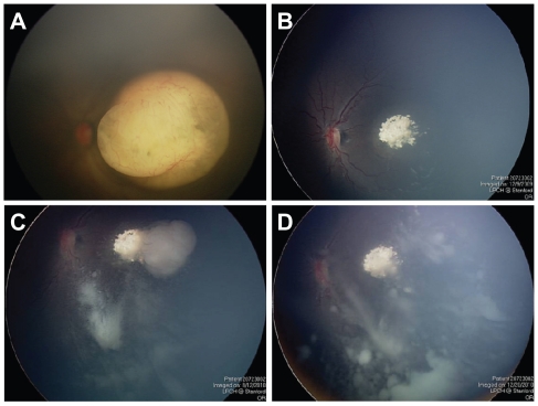
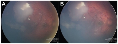
Selective intra-arterial chemotherapy (IAC) has been adopted by many ocular oncology centers to treat advanced intraocular retinoblastoma. In this report, we describe two patients with unilateral intraocular retinoblastoma and persistent vitreous seeding, who were treated with IAC after failed systemic chemotherapy. Despite multiple sessions and increasing dosage of drug delivery, vitreous seeding in these cases failed to respond to IAC, and ultimately both eyes were enucleated for tumor control. Based on the histopathologic findings in these two cases, IAC appears to have limitations in treating persistent vitreous seeding in eyes which have failed systemic chemotherapy. Possible causes for failure of IAC to treat persistent vitreous seeding include poor vitreous penetration, inactive state of tumor seeds within the avascular vitreous cavity, and chemotherapeutic drug resistance.
Keywords: chemotherapy; enucleation; eye; failure; intra-arterial; retinoblastoma.
Figures


References
-
- Abramson DH, Dunkel IJ, Brodie SE, Kim JW, Gobin YP. A phase I/II study of direct intraarterial (ophthalmic artery) chemotherapy with melphalan for intraocular retinoblastoma initial results. O–hthalmology. 2008;115(8):1398–1404. - PubMed
-
- Gobin YP, Dunkel IJ, Marr BP, Brodie SE, Abramson DH. Intra-arterial chemotherapy for the management of retinoblastoma: Four-year experience. Arch Ophthalmol. 2011;129(6):732–737. - PubMed
-
- Peterson EC, Elhammady MS, Quintero-Wolfe S, Murray TG, Aziz-Sultan MA. Selective ophthalmic artery infusion of chemotherapy for advanced intraocular retinoblastoma: initial experience with 17 tumors. J Neurosurg. 2011;114(6):1603–1608. - PubMed
-
- Shields CL, Ramasubramanian A, Rosenwasser R, Shields JA. Superselective catheterization of the ophthalmic artery for intraarterial chemotherapy for retinoblastoma. Retina. 2009;29(8):1207–1209. - PubMed
-
- Gombos DS, Hungerford J, Abramson DH, et al. Secondary acute myelogenous leukemia in patients with retinoblastoma: Is chemotherapy a factor? Ophthalmology. 2007;114(7):1378–1383. - PubMed
Publication types
LinkOut - more resources
Full Text Sources
