. 2011 Dec;7(12):e1002422.
doi: 10.1371/journal.ppat.1002422. Epub 2011 Dec 8.

IRGM is a common target of RNA viruses that subvert the autophagy network

Affiliations

IRGM is a common target of RNA viruses that subvert the autophagy network

Isabel Pombo Grégoire et al. PLoS Pathog. 2011 Dec.

Abstract

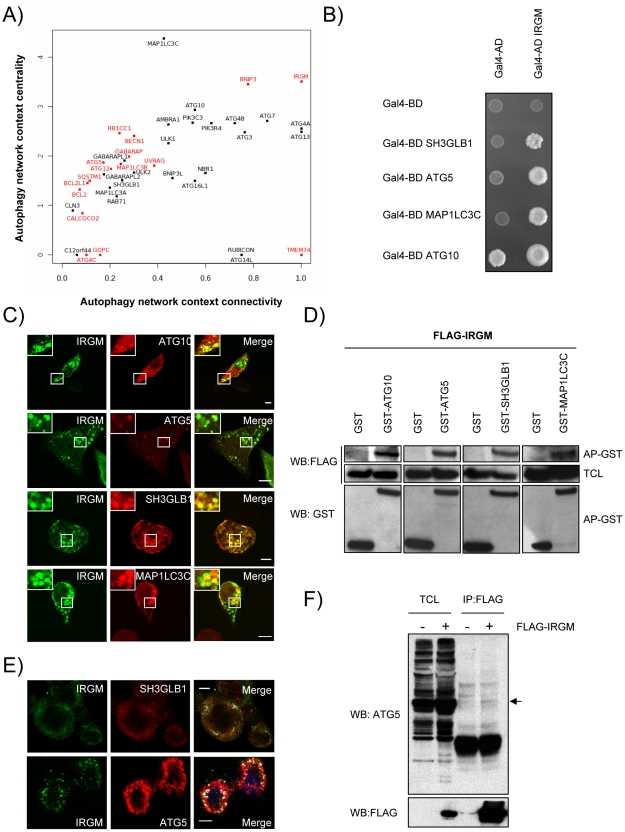
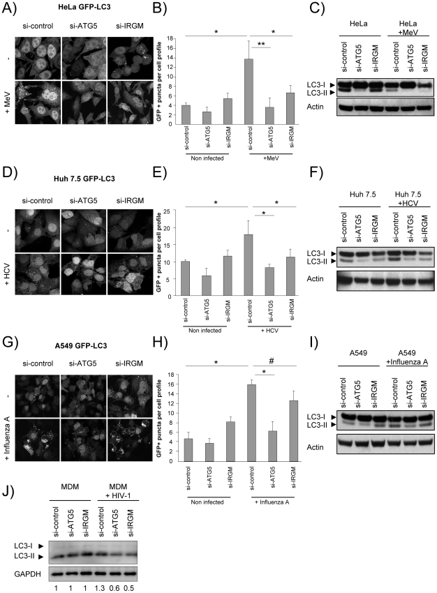
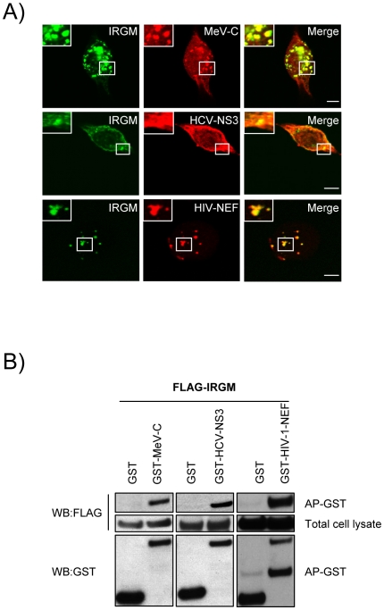
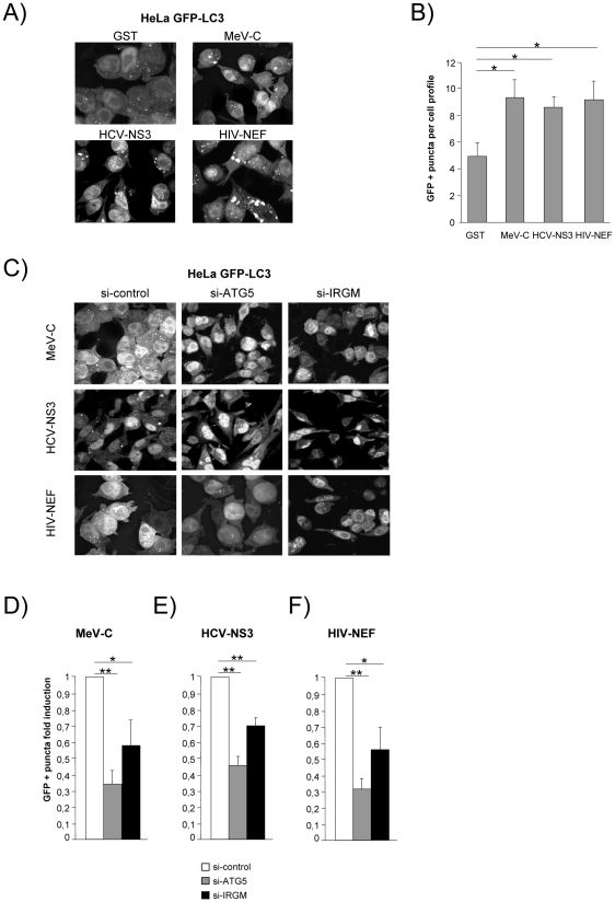
Autophagy is a conserved degradative pathway used as a host defense mechanism against intracellular pathogens. However, several viruses can evade or subvert autophagy to insure their own replication. Nevertheless, the molecular details of viral interaction with autophagy remain largely unknown. We have determined the ability of 83 proteins of several families of RNA viruses (Paramyxoviridae, Flaviviridae, Orthomyxoviridae, Retroviridae and Togaviridae), to interact with 44 human autophagy-associated proteins using yeast two-hybrid and bioinformatic analysis. We found that the autophagy network is highly targeted by RNA viruses. Although central to autophagy, targeted proteins have also a high number of connections with proteins of other cellular functions. Interestingly, immunity-associated GTPase family M (IRGM), the most targeted protein, was found to interact with the autophagy-associated proteins ATG5, ATG10, MAP1CL3C and SH3GLB1. Strikingly, reduction of IRGM expression using small interfering RNA impairs both Measles virus (MeV), Hepatitis C virus (HCV) and human immunodeficiency virus-1 (HIV-1)-induced autophagy and viral particle production. Moreover we found that the expression of IRGM-interacting MeV-C, HCV-NS3 or HIV-NEF proteins per se is sufficient to induce autophagy, through an IRGM dependent pathway. Our work reveals an unexpected role of IRGM in virus-induced autophagy and suggests that several different families of RNA viruses may use common strategies to manipulate autophagy to improve viral infectivity.


Conflict of interest statement

The authors have declared that no competing interests exist.

Figures

Figure 1
Figure 1. Targeting of the autophagy network by RNA viruses.
(A) List of the 44 autophagy-associated proteins considered in this study. Proteins represented in blue were used in yeast two-hybrid arrays. (B) Autophagy-associated proteins are targeted by RNA viruses. Graphical representation of ppi network of autophagy proteins and RNA virus proteins. Red nodes represent autophagy-associated proteins. Dark grey nodes represent RNA viral families. Blue edges represent novel ppi and gray edges represent ppi retrieved from the literature. The width of the red nodes is proportional to the number of viral families targeting the autophagy-associated proteins. (C) RNA viruses targeted human autophagy network. Graphical representation of the targeted human autophagy network. Each node represents one autophagy-associated protein. Black nodes are autophagy-associated proteins with unknown RNA virus interactions and red nodes represent autophagy-associated proteins targeted by at least one RNA virus protein. Blue edges represent novel ppi and gray edges represent ppi retrieved from the literature. The width of the nodes is proportional to the number of autophagy-associated proteins directly interacting with the considered protein.
Figure 2
Figure 2. IRGM, a protein that is highly targeted by RNA viruses, co-localizes and interacts with several autophagy-associated proteins.
(A) Autophagy-associated proteins contribute differently to the autophagy network. Autophagy-associated proteins are plotted according to their context connectivity and context centrality. Autophagy context connectivity (x axis) is the ratio of interaction in autophagy network over the interaction in the whole human protein interaction network. Autophagy context centrality (y axis) is the ratio of betweenness (log normalized values) in autophagy network over the betweenness in the human protein interaction network. A protein with high degree and high betweenness ratios is respectively defined as highly devoted and highly central in the autophagy context. Proteins in red represent autophagy-associated proteins targeted by at least one RNA virus protein. The four proteins that are not connected to the autophagy network are not represented. (B) IRGM interacts with ATG10, ATG5, SH3GLB1 and MAP1LC3C. IRGM was tested by yeast two-hybrid against 35 different autophagy-associated proteins. Positive interactions with ATG10, ATG5, SH3GLB1 and MAP1LC3C were found. A reduced yeast two-hybrid matrix containing positive interactions and the appropriate empty vector controls is shown. One experiment representative of three is shown. (C) IRGM co-localizes with autophagy-associated proteins. GFP-IRGM was expressed in HeLa cells together with FLAG-ATG10, FLAG-ATG5, FLAG-SH3GLB1 or FLAG-MAP1LC3C. Fixed cells were then stained with an anti-FLAG antibody and GFP-IRGM and FLAG-tagged proteins co-localisation was visualized on merged images by confocal microscopy. Scale bars, 5 µM. (D) IRGM interacts with autophagy-associated proteins. HEK293T cells were co-transfected with GST and GST-tagged expression vectors encoding the indicated proteins and FLAG-IRGM. Interaction was assayed by co-affinity purification (AP) using glutathione-sepharose beads. FLAG-IRGM was detected using anti-FLAG antibody after (AP-GST, WB: FLAG) and before (total cell lysate-TCL, WB: FLAG) co-AP. GST alone and GST-tagged proteins were detected by using anti-GST antibody (AP-GST, WB: GST). One experiment representative of two is shown. (E) Endogenous IRGM co-localizes with endogenous SH3GLB1 and ATG5 in MeV infected cells. HeLa cells were infected with MeV Edmonston (MOI = 1) for 24 hrs. Cells were fixed in acetone and both IRGM and/or SH3GLB1 or ATG5 were detected using specific antibodies. IRGM/SH3GLB1 or IRGM/ATG5 protein co-localisation was visualized on merged images obtained by confocal microscopy. Scale bars, 5 µM. (F) Endogenous ATG5 interacts with FLAG-IRGM. HeLa cells were transfected or not with FLAG-IRGM encoding vector and infected with MeV (MOI = 1) 24 hrs post-transfection. Cells were lysed 24 hrs post-infection. Flag-tagged IRGM was immunoprecipitated and endogenous ATG5 binding was detected by western blot (top panel). Overexpression and immunoprecipitation of FLAG tagged IRGM was confirmed by a western blot using (bottom panel).
Figure 3
Figure 3. MeV, HCV and HIV-1-induced autophagosome accumulation is dependent on IRGM.
(A–C) MeV-induced autophagy is dependent on IRGM. GFP-LC3-HeLa (A–B) or HeLa cells (C) were treated with si-control, si-ATG5 or si-IRGM and either left uninfected or infected with MeV Edmonston (MOI = 3) for 24 hrs. Autophagy was monitored either by evaluating the number of GFP-LC3+ vesicles per cell profile by confocal microscopy (A, B) or by detection of LC3-I and LC3-II by western-blot (C). Representative profiles for each condition (A) and the corresponding graph representing the number of GFP-LC3+ vesicles per cell profile (B) are shown, error bars, mean ± SD of three independent experiments. (C) One experiment representative of three is shown. (D–F) HCV-induced autophagy is dependent on IRGM. GFP-LC3-Huh7.5 (D–E) or Huh7.5 (F) cells were treated with the indicated si-RNA and either left uninfected or infected with HCV JFH-1 (MOI = 1) for 24 hrs. Autophagy was monitored as above. (E) Error bars, mean ± SD of three independent experiments. (F) One experiment representative of two is shown. (G–I) Influenza A-induced autophagy is not impaired by IRGM absence. GFP-LC3-A549 (G–H) or A549 (I) cells were treated with the indicated si-RNA and either left uninfected or infected with influenza A/H1N1/New Caledonia (MOI = 1) for 24 hrs. Autophagy was monitored as above. (H) Error bars, mean ± SD of two independent experiments. (I) One experiment representative of two is shown. (J) HIV-1-induced autophagy is dependent on IRGM. Monocyte-derived macrophages (MDM) were treated with the indicated si-RNA and were either left uninfected or infected with HIV-1 for 24 hrs. LC3-I and LC3-II detection was carried out by western-blot as in C, F, I. One experiment representative of two is shown with the quantification number representing the intensity of LC3-II/GAPDH bands normalized to the uninfected condition. Student's t test; * p<0.05; ** p<0.01; # p>0.05.
Figure 4
Figure 4. IRGM reduced expression efficiently decreases MeV, HCV and HIV-1 viral particles production.
(A) IRGM is necessary for MeV infectious particle production. HeLa cells were treated with the indicated si-RNAs. Cells were infected with MeV (MOI = 3) for 24 or 48 hrs. Results display average total infectious titers expressed as plate-forming units (pfu)/ml. Error bars, mean ± SD of three independent experiments. (B) IRGM is required for HCV JFH-1 infectious particle production. Huh-7.5 cells were treated with the indicated si-RNAs prior to cell infection with HCV JFH-1 (MOI = 1). The infectivity of the virus-containing cell supernatants was determined at 24 and 48 hrs post-infection. Results display average infectious titers. Error bars, mean ± SD of respectively three (24 hrs) or two (48 hrs) independent experiments. (C) IRGM-reduced expression does not modulate influenza A infectious particle production. A549 cells were treated with the indicated si-RNAs prior to cell infection with Influenza A/H1N1/New Caledonia (MOI = 0.1). The infectivity of the virus-containing cell supernatants was determined at 24 hrs post-infection. Results display average infectious titers. Error bars, mean ± SD of two independent experiments. (D) IRGM-reduced expression modulates HIV-1 production. MDM were treated with the indicated si-RNAs prior to cell infection with HIV-1. The infectivity was determined at 24 hrs post-infection. Results correspond to one experiment in triplicate, representative of four independent experiments. Student's t test; * p<0.05; ** p<0.01; # p>0.05.
Figure 5
Figure 5. Co-localisation and physical interaction of IRGM with MeV-C, HCV-NS3 and HIV-NEF proteins.
(A) IRGM co-localises with MeV-C, HCV-NS3 and HIV-NEF. GFP-IRGM was expressed in HeLa cells together with FLAG-MeV-C or FLAG-HCV-NS3 or FLAG-HIV-NEF. Co-localisation between GFP-IRGM and FLAG-tagged proteins was visualized as in figure 2B. Scale bars, 5 µM. (B) IRGM physically interacts with MeV-C, HCV-NS3 and HIV-NEF. HEK-293T cells were co-transfected with expression vectors encoding FLAG-IRGM and GST alone or GST fused to the indicated viral proteins. Co-purification of IRGM with viral proteins was assessed as in figure 2C. One experiment representative of two is shown.
Figure 6
Figure 6. MeV-C, HCV-NS3 and HIV-NEF proteins modulate autophagy via IRGM.
(A–B) Overexpression of MeV-C, HCV-NS3 and HIV-NEF modulates autophagosome formation. (A–B) GFP-LC3 HeLa cells were transfected with a GST encoding vector (control) or a vector encoding MeV-C, HCV-NS3 and HIV-NEF proteins. Twenty four hours post transfection the number of autophagic vesicles was determined by confocal microscopy. Representative profiles for each condition (A) and the corresponding graph representing the number of GFP-LC3+ vesicles per cell profile (B) are shown, error bars, mean ± SD of three independent experiments. (C–F) MeV-C, HCV-NS3 and HIV-NEF modulate autophagosome formation partly via IRGM. (C) GFP-LC3 HeLa cells were treated with si-control, si-ATG5 or si-IRGM 24 hrs prior transfection with vector encoding for MeV-C, HCV-NS3 or HIV-NEF. After an additional 24 hrs, cells were fixed and the number of autophagosome was determined by confocal microscopy. Representative profiles for each condition (C) and the corresponding graph representing the number of GFP-LC3+ vesicles per cell profile (D–F) are shown, error bars, mean ± SD of three independent experiments. Student's t test; * p<0.05; ** p<0.01.

Comment in

References

    1. Xie Z, Klionsky DJ. Autophagosome formation: core machinery and adaptations. Nat Cell Biol. 2007;9:1102–1109. - PubMed
    1. Levine B, Klionsky DJ. Development by self-digestion: molecular mechanisms and biological functions of autophagy. Dev Cell. 2004;6:463–477. - PubMed
    1. Furuya N, Yu J, Byfield M, Pattingre S, Levine B. The evolutionarily conserved domain of Beclin 1 is required for Vps34 binding, autophagy and tumor suppressor function. Autophagy. 2005;1:46–52. - PubMed
    1. Kihara A, Kabeya Y, Ohsumi Y, Yoshimori T. Beclin-phosphatidylinositol 3-kinase complex functions at the trans-Golgi network. EMBO Rep. 2001;2:330–335. - PMC - PubMed
    1. Zeng X, Overmeyer JH, Maltese WA. Functional specificity of the mammalian Beclin-Vps34 PI 3-kinase complex in macroautophagy versus endocytosis and lysosomal enzyme trafficking. J Cell Sci. 2006;119:259–270. - PubMed

Publication types

MeSH terms

LinkOut - more resources