

The human cytomegalovirus UL11 protein interacts with the receptor tyrosine phosphatase CD45, resulting in functional paralysis of T cells
- PMID: 22174689
- PMCID: PMC3234252
- DOI: 10.1371/journal.ppat.1002432
The human cytomegalovirus UL11 protein interacts with the receptor tyrosine phosphatase CD45, resulting in functional paralysis of T cells
Abstract
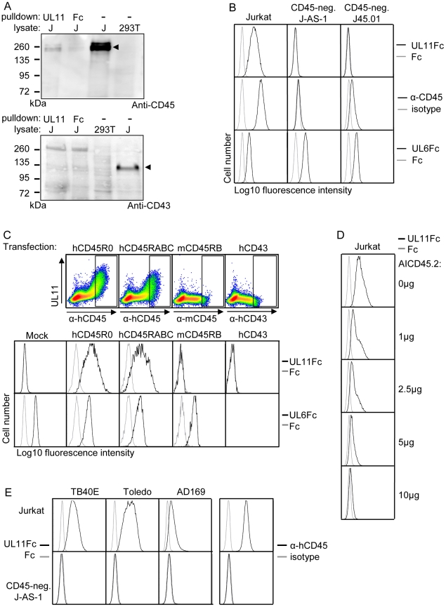
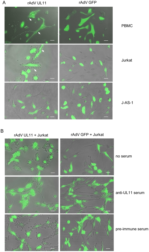
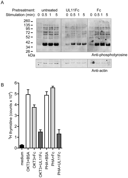
Human cytomegalovirus (CMV) exerts diverse and complex effects on the immune system, not all of which have been attributed to viral genes. Acute CMV infection results in transient restrictions in T cell proliferative ability, which can impair the control of the virus and increase the risk of secondary infections in patients with weakened or immature immune systems. In a search for new immunomodulatory proteins, we investigated the UL11 protein, a member of the CMV RL11 family. This protein family is defined by the RL11 domain, which has homology to immunoglobulin domains and adenoviral immunomodulatory proteins. We show that pUL11 is expressed on the cell surface and induces intercellular interactions with leukocytes. This was demonstrated to be due to the interaction of pUL11 with the receptor tyrosine phosphatase CD45, identified by mass spectrometry analysis of pUL11-associated proteins. CD45 expression is sufficient to mediate the interaction with pUL11 and is required for pUL11 binding to T cells, indicating that pUL11 is a specific CD45 ligand. CD45 has a pivotal function regulating T cell signaling thresholds; in its absence, the Src family kinase Lck is inactive and signaling through the T cell receptor (TCR) is therefore shut off. In the presence of pUL11, several CD45-mediated functions were inhibited. The induction of tyrosine phosphorylation of multiple signaling proteins upon TCR stimulation was reduced and T cell proliferation was impaired. We therefore conclude that pUL11 has immunosuppressive properties, and that disruption of T cell function via inhibition of CD45 is a previously unknown immunomodulatory strategy of CMV.
Conflict of interest statement
The authors have declared that no competing interests exist.
Figures
Similar articles
-
The human cytomegalovirus glycoprotein pUL11 acts via CD45 to induce T cell IL-10 secretion.PLoS Pathog. 2017 Jun 19;13(6):e1006454. doi: 10.1371/journal.ppat.1006454. eCollection 2017 Jun. PLoS Pathog. 2017. PMID: 28628650 Free PMC article.
-
Human Cytomegalovirus pUL11, a CD45 Ligand, Disrupts CD4 T Cell Control of Viral Spread in Epithelial Cells.mBio. 2022 Dec 20;13(6):e0294622. doi: 10.1128/mbio.02946-22. Epub 2022 Nov 29. mBio. 2022. PMID: 36445084 Free PMC article.
-
Expression of the human cytomegalovirus UL11 glycoprotein in viral infection and evaluation of its effect on virus-specific CD8 T cells.J Virol. 2014 Dec;88(24):14326-39. doi: 10.1128/JVI.01691-14. Epub 2014 Oct 1. J Virol. 2014. PMID: 25275132 Free PMC article.
-
CD45 in human physiology and clinical medicine.Immunol Lett. 2018 Apr;196:22-32. doi: 10.1016/j.imlet.2018.01.009. Epub 2018 Jan 31. Immunol Lett. 2018. PMID: 29366662 Review.
-
Refining human T-cell immunotherapy of cytomegalovirus disease: a mouse model with 'humanized' antigen presentation as a new preclinical study tool.Med Microbiol Immunol. 2016 Dec;205(6):549-561. doi: 10.1007/s00430-016-0471-0. Epub 2016 Aug 18. Med Microbiol Immunol. 2016. PMID: 27539576 Review.
Cited by
-
Evolution of the Cytomegalovirus RL11 gene family in Old World monkeys and Great Apes.Virus Evol. 2024 Aug 24;10(1):veae066. doi: 10.1093/ve/veae066. eCollection 2024. Virus Evol. 2024. PMID: 39315401 Free PMC article.
-
Identification and Functional Characterization of a Novel Fc Gamma-Binding Glycoprotein in Rhesus Cytomegalovirus.J Virol. 2019 Feb 5;93(4):e02077-18. doi: 10.1128/JVI.02077-18. Print 2019 Feb 15. J Virol. 2019. PMID: 30487278 Free PMC article.
-
The extracellular interactome of the human adenovirus family reveals diverse strategies for immunomodulation.Nat Commun. 2016 May 5;7:11473. doi: 10.1038/ncomms11473. Nat Commun. 2016. PMID: 27145901 Free PMC article.
-
A Prominent Role of the Human Cytomegalovirus UL8 Glycoprotein in Restraining Proinflammatory Cytokine Production by Myeloid Cells at Late Times during Infection.J Virol. 2018 Apr 13;92(9):e02229-17. doi: 10.1128/JVI.02229-17. Print 2018 May 1. J Virol. 2018. PMID: 29467314 Free PMC article.
-
Expression of viral CD45 ligand E3/49K on porcine cells reduces human anti-pig immune responses.Sci Rep. 2023 Oct 11;13(1):17218. doi: 10.1038/s41598-023-44316-y. Sci Rep. 2023. PMID: 37821577 Free PMC article.
References
-
- Mocarski E, Shenk T, Pass R. Cytomegaloviruses. In: Knipe DM, Howley PM, editors. Fields' Virology 5th Edition. Philadelphia (PA): Lippincott, Williams and Wilkins; 2007. pp. 2702–2772.
-
- Jackson SE, Mason GM, Wills MR. Human cytomegalovirus immunity and immune evasion. Virus Res. 2011;157:151–160. - PubMed
-
- Görzer I, Kerschner H, Redlberger-Fritz M, Puchhammer-Stöckl E. Human cytomegalovirus (HCMV) genotype populations in immunocompetent individuals during primary HCMV infection. J Clin Virol. 2010;48:100–103. - PubMed
-
- Meyer-König U, Ebert K, Schrage B, Pollak S, Hufert FT. Simultaneous infection of healthy people with multiple human cytomegalovirus strains. Lancet. 1998;352:1280–1281. - PubMed
Publication types
MeSH terms
Substances
LinkOut - more resources
Full Text Sources
Other Literature Sources
Medical
Molecular Biology Databases
Research Materials
Miscellaneous
