. 2011;6(12):e28015.
doi: 10.1371/journal.pone.0028015. Epub 2011 Dec 9.

Characterization of pellicle inhibition in Gluconacetobacter xylinus 53582 by a small molecule, pellicin, identified by a chemical genetics screen

Affiliations

Characterization of pellicle inhibition in Gluconacetobacter xylinus 53582 by a small molecule, pellicin, identified by a chemical genetics screen

Janice L Strap et al. PLoS One. 2011.

Abstract

Pellicin ([2E]-3-phenyl-1-[2,3,4,5-tetrahydro-1,6-benzodioxocin-8-yl]prop-2-en-1-one) was identified in a chemical genetics screen of 10,000 small molecules for its ability to completely abolish pellicle production in Gluconacetobacter xylinus. Cells grown in the presence of pellicin grew 1.5 times faster than untreated cells. Interestingly, growth in pellicin also caused G. xylinus cells to elongate. Measurement of cellulose synthesis in vitro showed that cellulose synthase activity was not directly inhibited by pellicin. Rather, when cellulose synthase activity was measured in cells that were pre-treated with the compound, the rate of cellulose synthesis increased eight-fold over that observed for untreated cells. This phenomenon was also apparent in the rapid production of cellulose when cells grown in the presence of pellicin were washed and transferred to media lacking the inhibitor. The rate at which cellulose was produced could not be accounted for by growth of the organism. Pellicin was not detected when intracellular contents were analyzed. Furthermore, it was found that pellicin exerts its effect extracellularly by interfering with the crystallization of pre-cellulosic tactoidal aggregates. This interference of the crystallization process resulted in enhanced production of cellulose II as evidenced by the ratio of acid insoluble to acid soluble product in in vitro assays and confirmed in vivo by scanning electron microscopy and powder X-ray diffraction. The relative crystallinity index, RCI, of pellicle produced by untreated G. xylinus cultures was 70% while pellicin-grown cultures had RCI of 38%. Mercerized pellicle of untreated cells had RCI of 42%, which further confirms the mechanism of action of pellicin as an inhibitor of the cellulose I crystallization process. Pellicin is a useful tool for the study of cellulose biosynthesis in G. xylinus.


Conflict of interest statement

Competing Interests: The authors have declared that no competing interests exist.

Figures

Figure 1
Figure 1. Chemical structure of pellicin.
Figure 2
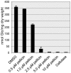
Figure 2. Dose-response curve for pellicin inhibition of G. xylinus pellicle.
In each case G. xylinus cultures were grown from a starting culture with an OD600 = 0.15 and either DMSO or pellicin was added to the culture and allowed to grow without shaking for 2 days at 30°C. Cellulase was added to 0.1% (w/v). Values are averages ± SD; n = 3.
Figure 3
Figure 3. The influence of pellicin on G. xylinus growth.
Pellicin does not affect the viability of G. xylinus. G. xylinus was grown at 30°C in Schramm-Hestrin broth containing 0.1% cellulase and either 10 µM pellicin (▪) or DMSO (▴) as a control. Data show the mean ± SD of four experimental determinations.
Figure 4
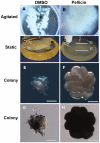
Figure 4. Effect of pellicin on cellulose production in liquid culture and solid medium.
G. xylinus was grown at 30°C in liquid culture under agitated (A, B) and static conditions (C, D); (A, C) are DMSO controls, (B, D) grown in the presence of 10 µM pellicin. Arrows in (A, C) indicate either the large aggregates or pellicle that form in the absence of pellicin. Colony morphology of G. xylinus grown on SH agar plates that were supplemented with (E, G) DMSO or (F, H) 10 µM pellicin. Photographs of (E, F) were taken with illumination from above and (G, H) were taken with illumination from below of the same colonies. Note the larger, undulate, raised colonies forming on pellicin supplemented plates (F). Arrows in (G) indicate the filiform projections emerging from colony, which are absent in (H). Scale bars equal 0.5 mm.
Figure 5
Figure 5. Pellicin affects the cell length of G. xylinus.
Box plot of cell lengths of G. xylinus grown in the presence and absence of pellicin. The box signifies the upper and lower quartiles for each growth condition and the median is represented by the black line within each box. Outliers are denoted by (○). Extreme outliers are denoted by (*). Cell lengths of 500 cells from each growth condition were measured (p<0.001, independent samples t-test).
Figure 6
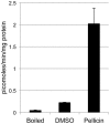
Figure 6. In vitro cellulose synthase assay using membrane preparations of G. xylinus grown in the presence and absence of pellicin using UDP-[3H]glucose as substrate.
1) Boiled control 2) Untreated 3) Pellicin pretreated. Cells grown in the presence of pellicin show an increased cellulose synthase activity. Data show the mean ± SE of three experimental determinations.
Figure 7
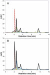
Figure 7. Pellicin remains in the extracellular environment.
High performance liquid chromatography of organic extracts of A) G. xylinus in the absence and B) presence of pellicin. Data shown is representative of triplicate experiments: (red dashed line) pellicin standard, (black line) extracellular supernatant, (blue line) pellet wash prior to cellulase treatment, (green line) supernatant from cellulase treated cells, (orange line) cell free lysate, (purple line) cellular debris analyzed at 220 nm, the optimum wavelength for pellicin detection.
Figure 8
Figure 8. Pellicin affects the crystallinity of cellulose.
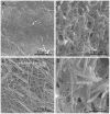
Scanning electron micrographs of G. xylinus culture grown in the SH medium containing DMSO (A and C) and 10 µM pellicin (B and D). Untreated cultures have distinct, long cellulose-I fibrils that surround G. xylinus cells (C), while cultures treated with pellicin have shortened, cellulose-II strands that appears to be more gel-like (D). Scale bar are 8.6 µm in A and B and 2.7 µm in C and D.
Figure 9
Figure 9. Pellicin affects the crystallization of cellulose produced by G. xylinus.
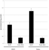
In vitro cellulose synthase assays of crude enzyme prepared from G. xylinus grown SH medium and incubated with DMSO or pellicin for 1 hour prior to UDP-[3H]glucose addition. Reactions treated with 0.5 M NaOH contain both cellulose I and cellulose II. Acetic-nitric acid treatment of reactions removes cellulose II while retaining cellulose I. Data show the mean ± SE of three experimental determinations.
Figure 10
Figure 10. Relative proportion of cellulose I to cellulose II produced during in vitro cellulose synthesis assay.
The ratio of cellulose I to cellulose II production was determined from crude enzyme preparations of G. xylinus grown in DMSO and 10 µM pellicin by assaying the amount of UDP-[3H]glucose in the acid insoluble and soluble fractions as described in the Materials and Methods.
Figure 11
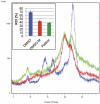
Figure 11. Powder X-ray diffraction pattern for extracellular cellulose produced by G. xylinus grown in the absence (blue) or presence of 10 µM pellicin (green) compared to mercerized bacterial cellulose (red).
Inset, relative crystallinity index (RCI) of cellulose produced under different conditions calculated from X-ray diffraction (see Materials and Methods for details). Values ± SD are shown; n = 3 for each treatment.

References

    1. Delmer D, Haigler C. The regulation of metabolic flux to cellulose, a major sink for carbon in plants. Metab Eng. 2002;4:22–28. - PubMed
    1. Fu GZ, Chan AW, Minns DE. Life cycle assessment of bio-ethanol derived from cellulose. Int J LCA. 2003;8:137–141.
    1. Shah J, Brown RM., Jr Towards electronic paper displays made from microbial cellulose. Appl Microbiol Biotechnol. 2005;66:352–355. - PubMed
    1. Czaja W, Krystynowicz A, Bielecki S, Brown R., Jr Microbial cellulose – the natural power to heal wounds. Biomaterials. 2006;27:145–151. - PubMed
    1. Ross P, Mayer R, Benziman M. Cellulose biosynthesis and function in bacteria. Microbiol Mol Biol Rev. 1991;55:35–58. - PMC - PubMed

Publication types

MeSH terms