. 2011;6(12):e28017.
doi: 10.1371/journal.pone.0028017. Epub 2011 Dec 8.

Role of collagens and perlecan in microvascular stability: exploring the mechanism of capillary vessel damage by snake venom metalloproteinases

Affiliations

Role of collagens and perlecan in microvascular stability: exploring the mechanism of capillary vessel damage by snake venom metalloproteinases

Teresa Escalante et al. PLoS One. 2011.

Abstract

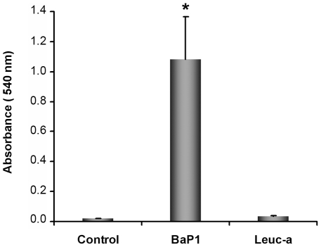
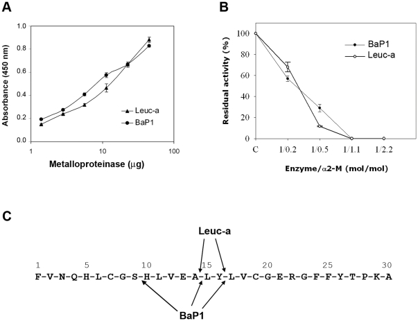
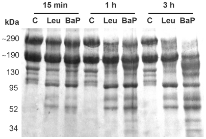
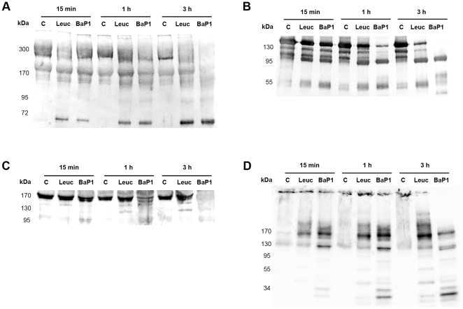
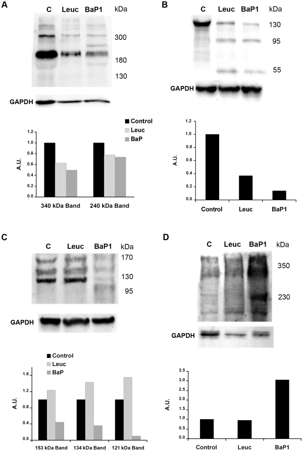
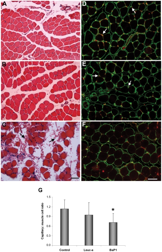
Hemorrhage is a clinically important manifestation of viperid snakebite envenomings, and is induced by snake venom metalloproteinases (SVMPs). Hemorrhagic and non-hemorrhagic SVMPs hydrolyze some basement membrane (BM) and associated extracellular matrix (ECM) proteins. Nevertheless, only hemorrhagic SVMPs are able to disrupt microvessels; the mechanisms behind this functional difference remain largely unknown. We compared the proteolytic activity of the hemorrhagic P-I SVMP BaP1, from the venom of Bothrops asper, and the non-hemorrhagic P-I SVMP leucurolysin-a (leuc-a), from the venom of Bothrops leucurus, on several substrates in vitro and in vivo, focusing on BM proteins. When incubated with Matrigel, a soluble extract of BM, both enzymes hydrolyzed laminin, nidogen and perlecan, albeit BaP1 did it at a faster rate. Type IV collagen was readily digested by BaP1 while leuc-a only induced a slight hydrolysis. Degradation of BM proteins in vivo was studied in mouse gastrocnemius muscle. Western blot analysis of muscle tissue homogenates showed a similar degradation of laminin chains by both enzymes, whereas nidogen was cleaved to a higher extent by BaP1, and perlecan and type IV collagen were readily digested by BaP1 but not by leuc-a. Immunohistochemistry of muscle tissue samples showed a decrease in the immunostaining of type IV collagen after injection of BaP1, but not by leuc-a. Proteomic analysis by LC/MS/MS of exudates collected from injected muscle revealed higher amounts of perlecan, and types VI and XV collagens, in exudates from BaP1-injected tissue. The differences in the hemorrhagic activity of these SVMPs could be explained by their variable ability to degrade key BM and associated ECM substrates in vivo, particularly perlecan and several non-fibrillar collagens, which play a mechanical stabilizing role in microvessel structure. These results underscore the key role played by these ECM components in the mechanical stability of microvessels.


Conflict of interest statement

Competing Interests: The authors have declared that no competing interests exist.

Figures

Figure 1
Figure 1. Hemorrhagic activity of BaP1 and leuc-a in mouse muscle.
Mice were injected intramuscularly, in the gastrocnemius, with 50 µg of either BaP1 or leuc-a, dissolved in 50 µL PBS, or with 50 µL PBS (controls). After 15 min, mice were sacrificed and muscles were dissected out and placed in 1.5 mL of distilled water. Samples were incubated overnight at 4°C and the absorbance at 540 nm was recorded in the supernatant as an indicator of muscle hemoglobin content. BaP1 induced a profuse hemorrhage whereas leuc-a did not exert hemorrhagic activity. Results are presented as mean ± SD (n = 4). *P<0.05 when compared to control and leuc-a treatments.
Figure 2
Figure 2. Proteolytic activity of BaP1 and leuc-a, and inhibition by α2-macroglobulin.
(A) Hydrolytic activity of BaP1 and leuc-a on azocasein. Various amounts of each enzyme were incubated with azocasein for 90 min at 37°C. The reaction was stopped by the addition of 5% trichloroacetic acid, and the absorbances of the supernatants at 450 nm were recorded after centrifugation. Controls of azocasein without enzyme were run in parallel and their absorbance was subtracted from the sample values. Results are presented as mean ± S.D. (n = 3). (B) Stoichiometry of inhibition of BaP1 and leuc-a by α2M. The plasma inhibitor was incubated with BaP1 or leuc-a at various molar ratios, and proteolytic activity was tested on dimethylcasein. The remaining protease activity is expressed as percentage of the original activity measured in the absence of the inhibitor. Results are presented as mean ± SD (n = 3). (C) Cleavage sites of BaP1 and leuc-a on oxidized insulin B-chain. After 30 min of digestion, peptides were separated by HPLC and identified by their amino acid sequence.
Figure 3
Figure 3. Hydrolysis of Matrigel proteins by BaP1 and leuc-a.
Matrigel was incubated at 37°C with each enzyme at a 1∶50 (w:w) enzyme:substrate ratio for 15 min, 1 h and 3 h. A control of Matrigel without enzymes (C) was included for each time interval. Matrigel solutions were separated by SDS–PAGE under reducing conditions using a 4–15% gradient gel, and transferred to nitrocellulose membrane and stained with Ponceau-S.
Figure 4
Figure 4. Hydrolysis of basement membrane components in vitro.
Hydrolysis of laminin (A), nidogen (B), type IV collagen (C) and perlecan (D) by BaP1 and leuc-a, as detected by Western blotting of Matrigel. Matrigel was incubated with either BaP1, leuc-a or PBS (control, lane C), as described in the legend of Fig 3. Matrigel preparations were separated by SDS-PAGE under reducing conditions using a 4-15% gradient gel and transferred to nitrocellulose membranes. Immunodetection was performed with either anti-laminin, anti-nidogen, anti-type IV collagen or anti-perlecan antibodies. Reaction was developed with a chemiluminiscent substrate.
Figure 5
Figure 5. Hydrolysis of basement membrane components in vivo.
Hydrolysis of laminin (A), nidogen (B), type IV collagen (C) and perlecan (D) by BaP1 and leuc-a, as detected by Western blotting of homogenates of injected mouse gastrocnemius muscle. Groups of mice were injected in the gastrocnemius muscle with either 50 µg BaP1, 50 µg leuc-a or PBS (lane C). After 15 min, mice were sacrificed and tissue was homogenized and centrifuged to obtain the supernatant. Supernatants of muscle homogenates were separated by SDS-PAGE under reducing conditions, using a 4–15% gradient gel, and transferred to nitrocellulose membranes. Immunodetection was performed with either anti-laminin, anti-nidogen, anti-type IV collagen or anti-endorepellin antibodies, and with anti-GAPDH as loading control in tissue homogenates. Reaction was developed with a chemiluminiscent substrate. Densitometry was carried out in blots of tissue homogenates with ImageLab software; a relative quantification was performed adjusting each sample to the corresponding control.
Figure 6
Figure 6. Histological and immunohistochemical analysis of the effects of BaP1 and leuc-a in skeletal muscle.
Mice were injected with either 50 µL PBS as control (A, D), 50 µg of leuc-a (B, E) or 50 µg BaP1 (C, F). Tissue samples were collected 15 min after injection and processed for embedding in paraffin (see experimental details in Methods). A, B and C: Hematoxylin-eosin staining. Hemorrhage, evidenced by the presence of erythrocytes in the interstitial space (arrows), occurred only in muscle injected with BaP1. D, E and F: Immunostaining with anti-type IV collagen (green) and anti-VEGFR2 (red). Arrows depict capillary vessels. There is a reduction in capillary vessels positive for type IV collagen and VEGFR2 in samples treated with BaP1, whereas no reduction in the staining of capillaries was observed in tissue injected with leuc-a. Bar corresponds to 50 µm. (G) The total number of capillaries and muscle cells were counted in various sections, and the capillary: muscle cell ratio was calculated. Results are presented as mean ± SD. A significant reduction in the ratio was observed only in muscle injected with BaP1, but not with leuc-a. *P<0.05 when compared with capillary: muscle cell ratios of control and leuc-a-treated samples.
Figure 7
Figure 7. SDS-PAGE of exudates samples collected from mice injected with either BaP1 or leuc-a.
Samples corresponding to 20 µg protein of exudates collected 15 min after injection of SVMPs were electrophoresed on a 4–20% gradient gel followed by staining with Coomassie Blue. Molecular mass markers are depicted to the left. Gel lanes were cut into ten equal size slices for further proteomic analysis (see Methods for details).

Similar articles

Cited by

References

    1. Fox JW, Serrano SMT. Structural considerations of the snake venom metalloproteinases, key members of the M12 reprolysin family of metalloproteinases. Toxicon. 2005;45:969–985. - PubMed
    1. Calvete JJ, Sanz L, Angulo Y, Lomonte B, Gutiérrez JM. Venoms, venomics, antivenomics. FEBS Lett. 2009;583:1736–1743. - PubMed
    1. Gutiérrez JM, Rucavado A, Escalante T. Snake venom metalloproteinases. Biological roles and participation in the pathophysiology of envenomation. In: Mackessy SP, editor. Handbook of Venoms and Toxins of Reptiles. Boca Raton: CRC Press; 2010. pp. 114–128.
    1. Gutiérrez JM, Rucavado A. Snake venom metalloproteinases: Their role in the pathogenesis of local tissue damage. Biochimie. 2000;82:841–850. - PubMed
    1. Kamiguti AS. Platelets as targets of snake venom metalloproteinases. Toxicon. 2005;45:1041–1049. - PubMed

Publication types

MeSH terms