Comparative Study
. 2011;6(12):e28429.
doi: 10.1371/journal.pone.0028429. Epub 2011 Dec 9.

Infection of differentiated porcine airway epithelial cells by influenza virus: differential susceptibility to infection by porcine and avian viruses

Affiliations
Comparative Study

Infection of differentiated porcine airway epithelial cells by influenza virus: differential susceptibility to infection by porcine and avian viruses

Darsaniya Punyadarsaniya et al. PLoS One. 2011.

Abstract

Background: Swine are important hosts for influenza A viruses playing a crucial role in the epidemiology and interspecies transmission of these viruses. Respiratory epithelial cells are the primary target cells for influenza viruses.

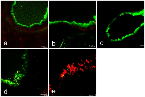
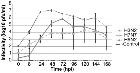
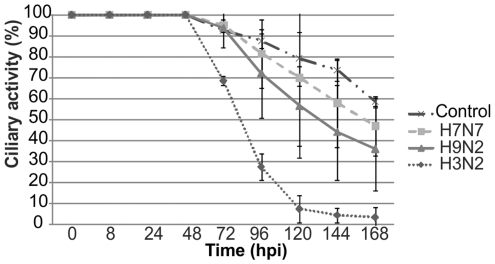
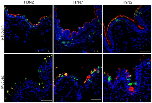
Methodology/principal findings: To analyze the infection of porcine airway epithelial cells by influenza viruses, we established precision-cut lung slices as a culture system for differentiated respiratory epithelial cells. Both ciliated and mucus-producing cells were found to be susceptible to infection by swine influenza A virus (H3N2 subtype) with high titers of infectious virus released into the supernatant already one day after infection. By comparison, growth of two avian influenza viruses (subtypes H9N2 and H7N7) was delayed by about 24 h. The two avian viruses differed both in the spectrum of susceptible cells and in the efficiency of replication. As the H9N2 virus grew to titers that were only tenfold lower than that of a porcine H3N2 virus this avian virus is an interesting candidate for interspecies transmission. Lectin staining indicated the presence of both α-2,3- and α-2,6-linked sialic acids on airway epithelial cells. However, their distribution did not correlate with pattern of virus infection indicating that staining by plant lectins is not a reliable indicator for the presence of cellular receptors for influenza viruses.

Conclusions/significance: Differentiated respiratory epithelial cells significantly differ in their susceptibility to infection by avian influenza viruses. We expect that the newly described precision-cut lung slices from the swine lung are an interesting culture system to analyze the infection of differentiated respiratory epithelial cells by different pathogens (viral, bacterial and parasitic ones) of swine.


Conflict of interest statement

Competing Interests: The authors have declared that no competing interests exist.

Figures

Figure 1
Figure 1. Vitality of PCLS evaluated by live (green)/dead (red) staining.
Slices were stained with a commercial kit at day 1, 3 and 7 after preparation (upper panels, a–c). For comparison, in the lower panels the live/dead staining is shown for two slices that either had retained full ciliary activity (100%) (d), or had completely lost ciliary activity (0%) (e). The scale bar indicates 50 µm.
Figure 2
Figure 2. Vitality of PCLS evaluated by bronchoconstriction.
To induce bronchoconstriction, the untreated slice (a) was incubated with 10−4 M methacholine (b). Removal of the drug resulted in a reverse effect (c).
Figure 3
Figure 3. Expression of sialic acid in swine PCLS cells.
Sialic acids were detected by lectin staining: MAA (Maackia amurensis agglutinin) for α2,3-linked sialic acids and SNA (Sambucus nigra agglutinin) for α2,6-linked sialic acids. Ciliated cells were stained using an anti-β-tubulin antibody and mucus-producing cells were stained using an anti muc5ac antibody. In panels A, lectin-staining is compared with staining of ciliated (red) or mucus-producing cells (red in Ab, green in Ad); SNA staining is shown in green (Aa and Ab), MAA staining is shown in green (Ac) or red (Ad). In Ba, co-staining of ciliated (red) and mucus-producing cells (green) is shown; Bb shows co-staining with MAA (red) and SNA (green). In C samples were co-stained SNA (green) and MAA (red); cryosections were derived from PCLS before (Ca) or after neuraminidase treatment (Cb).
Figure 4
Figure 4. Glycan array analysis.
Avian influenza virus (H9N2 and H7N7 subtypes) were subjected to glycan array analysis.
Figure 5
Figure 5. Infection of PCLS by porcine and avian influenza viruses evaluated by titration of infectious virus.
PCLS were mock-infected or infected by porcine H3N2, avian H9N2, or avian H7N7 virus. Up to 7 days post infection, infectious virus released into the supernatants of PCLS was titrated at daily intervals by plaque assays.
Figure 6
Figure 6. Infection of swine PCLS by porcine and avian influenza viruses evaluated by ciliary activity.
PCLS were mock-infected or infected by porcine H3N2, avian H9N2, or avian H7N7 virus. Up to seven days post infection, PCLS were analyzed for ciliary activity at daily intervals.
Figure 7
Figure 7. Infection of swine PCLS by influenza viruses characterized by immunostaining.
PCLS were infected by either porcine H3N2, avian H9N2, or avian H7N7 virus. Cryosections were prepared at 24 h.p.i. and used for detection of infected cells, ciliated cells, and mucus-producing cells. Infected cells were stained with an anti-nucleoprotein antibody (green); ciliated cells were stained using an anti-β-tubulin antibody (red) and mucus-producing cells were stained using an Muc5Ac antibody (red).

Similar articles

Cited by

References

    1. Guan Y, Shortridge KF, Krauss S, Li PH, Kawaoka Y, et al. Emergence of avian H1N1 influenza viruses in pigs in China. J Virol. 1996;70:8041–8046. - PMC - PubMed
    1. Karasin AI, Brown IH, Carman S, Olsen CW. Isolation and characterization of H4N6 avian influenza viruses from pigs with pneumonia in Canada. J Virol. 2000;74:9322–9327. - PMC - PubMed
    1. Peiris JS, Guan Y, Markwell D, Ghose P, Webster RG, et al. Cocirculation of avian H9N2 and contemporary “human” H3N2 influenza A viruses in pigs in southeastern China: potential for genetic reassortment? J Virol. 2001;75:9679–9686. - PMC - PubMed
    1. De Vleeschauwer A, Atanasova K, Van Borm S, van den Berg T, Rasmussen TB, et al. Comparative pathogenesis of an avian H5N2 and a swine H1N1 influenza virus in pigs. PLoS One. 2009;4:e6662. - PMC - PubMed
    1. Scholtissek C, Bürger H, Kistner O, Shortridge KF. The nucleoprotein as a possible major factor in determining host specificity of influenza H3N2 viruses. Virology. 1985;147:287–294. - PubMed

Publication types

MeSH terms