. 2011;6(12):e28650.
doi: 10.1371/journal.pone.0028650. Epub 2011 Dec 13.

MicroRNA expression characterizes oligometastasis(es)

Affiliations

MicroRNA expression characterizes oligometastasis(es)

Yves A Lussier et al. PLoS One. 2011.

Abstract

Background: Cancer staging and treatment presumes a division into localized or metastatic disease. We proposed an intermediate state defined by ≤ 5 cumulative metastasis(es), termed oligometastases. In contrast to widespread polymetastases, oligometastatic patients may benefit from metastasis-directed local treatments. However, many patients who initially present with oligometastases progress to polymetastases. Predictors of progression could improve patient selection for metastasis-directed therapy.

Methods: Here, we identified patterns of microRNA expression of tumor samples from oligometastatic patients treated with high-dose radiotherapy.

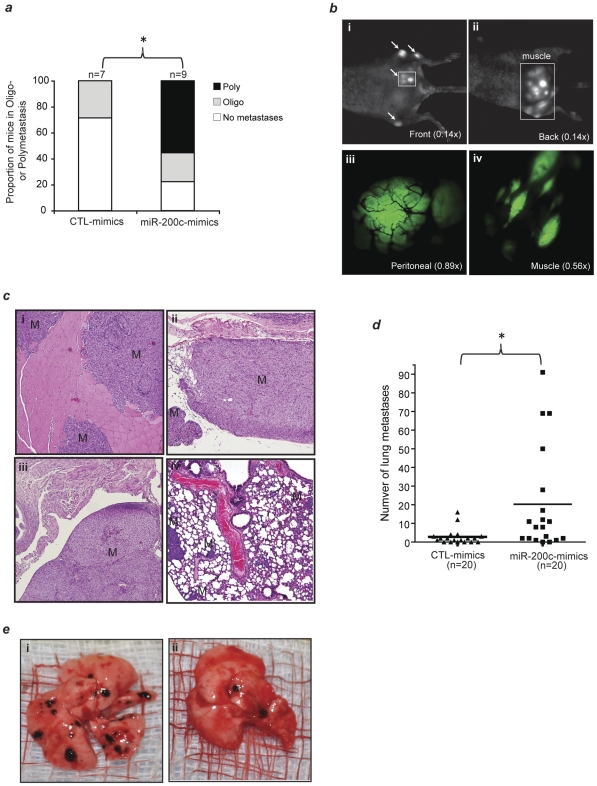
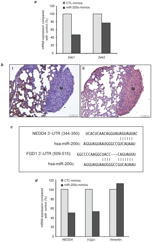
Results: Patients who failed to develop polymetastases are characterized by unique prioritized features of a microRNA classifier that includes the microRNA-200 family. We created an oligometastatic-polymetastatic xenograft model in which the patient-derived microRNAs discriminated between the two metastatic outcomes. MicroRNA-200c enhancement in an oligometastatic cell line resulted in polymetastatic progression.

Conclusions: These results demonstrate a biological basis for oligometastases and a potential for using microRNA expression to identify patients most likely to remain oligometastatic after metastasis-directed treatment.


Conflict of interest statement

Competing Interests: The authors have declared that no competing interests exist.

Figures

Figure 1
Figure 1. Unsupervised hierarchical clustering of:
(a) metastatic tumors microRNA expression showing clustering of oligo- vs polymetastatic samples. Red, black and green represent TaqMan qPCR Ct values above, at or below mean level, respectively, across all samples and 335 microRNAs. As shown, all seven polymetastatic samples are clustered together, while eight out of ten oligometastatic samples cluster together. This suggests that the oligo vs polymetastatic phenotype is overriding other predictable groupings such as histology of primary tumor and metastatic site. However, in the primary samples, the primary site was the dominant signal of the unsupervised hierarchical clustering (Fig. S1). (b) MicroRNA expression of five patients with paired primary and metastatic samples showing clustering of (i) primary (Pr) and metastasis(es) sample sites of the same patient and (ii) oligo (Ol-) vs polymetastatic (Pol-) progression phenotype across patients.
Figure 2
Figure 2. Validation of microRNA expression signatures in human datasets: prediction of oligometastatic progression by microRNA expression signatures.
The Receiver Operating Characteristic (ROC) curves describe how accurately the prioritized microRNAs can discriminate between oligo- vs poly- metastasis(es) samples by plotting the possible combinations of sensitivity and specificity obtained at different cutoff points of the prioritized microRNA classifier. (a) Pr-miRs, 17 prioritized microRNAs from the primary tumors sample ( Table 1b ), were used to predict oligometastasis(es) progression in the 16 metastatic tumor samples using permutation controlled ROC curves of the first PCA component (See Methods). (b) Similarly, M-miRs, 29 prioritized microRNAs from the metastatic tumor samples ( Table 1a ), were used to predict oligometastasis(es) progression in the 26 primary samples. Empirical P values of the AUC were calculated from empirical permutation resampling (see Methods S1).
Figure 3
Figure 3. Histological and in vivo characterization of oligo- and poly- metastasis(es) derived from tail-vein injected MDA-MB-435-GFP lung derivative cell lines.
2×10⁶ purified MDA-MB-435-GFP lung derivative cell lines established from lungs harboring oligo- (L1-R1) or poly-(L1Mic-R1) metastases respectively were injected via tail-vein. Animals developing macroscopic observable metastases were sacrificed at the time of this finding. The rest of the animals were sacrificed at 12-weeks post tumor cell injection. Necropsy was performed to score macroscopic metastatic lesions and lungs were harvested and paraffin embedded for histological characterization. (a) Representative lung metastatic-foci developed from oligmetastatic L1-R1 cell line harvested at week-12 or (b) a polymetastatic L1Mic-R1 cell line, harvested at week-7 shown by H&E staining (arrows, 40× magnification). (c) An enlargement (200×) of the insert in (b). (d) Representative fluorescent in vivo imaging identifying extensive lung and whole body polymetastatic lesions after tail vein injection with L1Mic-R1 cells (OV-100 imager, green fluorescence = metastatic lesions). (e) Oligo- vs polymetastases progression in these 29 NCI athymic female mice establish that polymetastatic L1Mic-R1 cells produced more aggressive metastatic progression than the oligometastatic L1-R1 cells (odds ratio at week 12 = 10; P = 0.0092; two-tailed Fischer Exact Test). Additionally, L1Mic-R1 produced more aggressive metastatic progression: at week 9, 73% of L1Mic-R1 had developed polymetastases as compared to none among those exposed to L1-R1 (P = 5×10⁻⁵; two-tailed Fischer Exact Test).
Figure 4
Figure 4. Validations of the prioritized human microRNAs in the animal model of oligo and polymetastases.
The prioritized microRNAs between oligometastatic and polymetastatic progression were identified in primary tumors and in metastatic tumors of clinical samples yielding two lists: Pr-miRs and M-miRs, respectively (see Table 1a–b ). These lists of microRNAs were used to rank the microRNA expression of seven cell line samples derived from animal modeling of oligometastasis(es) (L1-R1) and of widespread polymetastases (L1Mic-R1). MicroRNA expression was conducted in three oligometastatic L1-R2 lung cell lines as well as four polymetastatic L1Mic-R2 lung cell lines from seven distinct animals. Principal component analysis of the expression of microRNAs was conducted in these cell line samples without providing any information on the L1-R2 or L1Mic-R2 status. In each sample, the first component values of (a) Pr-miRs and of (b) M-miRs is sufficient to discriminate between the oligo- (L1) and polymetastatic (L1Mic) phenotype of the animal model (Pr-miRs P = 0.058; M-miRs P = 0.058; two-tailed Mann-Whitney U Test, Methods S1).
Figure 5
Figure 5. microRNA-200c regulate oligo- to poly- metastasis(es) progression in the L1-R2-435-GFP xenograft model.
2×10⁶ control-mimics or microRNA-200c specific mimics-treated L1-R2-435-GFP cells were tail-vein injected after 48 hr of transfection, and the development of macrometastases was monitored (Methods). (a) microRNA-200c mimics treatment significantly converted oligometastasis(es) to largely polymetastases. Poly: polymetastases; Oligo: oligometastasis(es). *P = 0.012 (one-tailed Mann Whitney U Test). (b) Non-invasive, variable magnification (0.14–0.89×) OV-100 fluorescent imaging visualization of polymetastatic dissemination in a representative animal injected with microRNA-200c mimics-treated L1-R2 cells. Arrows: macrometastases; green: L1-R2-435-GFP tumor; black lines in (iii): tumor blood vessels. (c) IHC confirmation of macrometastases in the muscle (i), peritoneum membrane (ii), peritoneal cavity (iii) and lung (iv). Magnification: 100×; M: macrometastases. (d) microRNA-200c mimics treatment significantly increased the efficiency of B16F1 mouse melanoma cells to form lung macrometastases. *P = 0.0057 (one-tailed Mann Whitney U Test). (e) Representative images of mouse lung obtained from animals tail vein-injected with microRNA-200c mimics treated (i) and control mimics treated (ii) B16F1 cells.
Figure 6
Figure 6. microRNA-200c mimics treatment lead to specific inhibition of its putative target gene expression.
L1-R2-435-GFP cells were treated with equal amount of control-mimics or microRNA-200c mimics for 48 hours (Method). Thereafter, one fifth of the transfected cells were used for total RNA extraction and the rest were used for tail-vein injection ( Figure 5 ). (a) TaqMan quantification of Zeb1 and Zeb2 mRNA expression. GPDH was used for normalization. (b) Lungs macrometastases derived from L1-R2-435-GFP cells treated with control mimics or microRNA-200c mimics were negative for E-cadherin (i) and positive for the EMT marker vimentin (ii). (c) TargetScan alignment of microRNA-200c binding site at 3′-UTR of two computationally prioritized microRNA-200c putative targets NEDD4 and FGD1. (d) TaqMan quantification of NEDD4, FGD1 and Vimentin mRNA expression. GPDH was used for normalization.

References

    1. Weichselbaum RR, Hellman S. Oligometastases revisited. Nat Rev Clin Oncol 2011 - PubMed
    1. Staren ED, Salerno C, Rongione A, Witt TR, Faber LP. Pulmonary resection for metastatic breast cancer. Arch Surg. 1992;127:1282–1284. - PubMed
    1. Hellman S, Weichselbaum RR. Oligometastases. J Clin Oncol. 1995;13:8–10. - PubMed
    1. Fong Y, Cohen AM, Fortner JG, Enker WE, Turnbull AD, et al. Liver resection for colorectal metastases. J Clin Oncol. 1997;15:938–946. - PubMed
    1. Tomlinson JS, Jarnagin WR, DeMatteo RP, Fong Y, Kornprat P, et al. Actual 10-year survival after resection of colorectal liver metastases defines cure. J Clin Oncol. 2007;25:4575–4580. - PubMed

Publication types

LinkOut - more resources