. 2011;6(12):e28753.
doi: 10.1371/journal.pone.0028753. Epub 2011 Dec 12.

An off-target nucleostemin RNAi inhibits growth in human glioblastoma-derived cancer stem cells

Affiliations

An off-target nucleostemin RNAi inhibits growth in human glioblastoma-derived cancer stem cells

Jon Gil-Ranedo et al. PLoS One. 2011.

Abstract

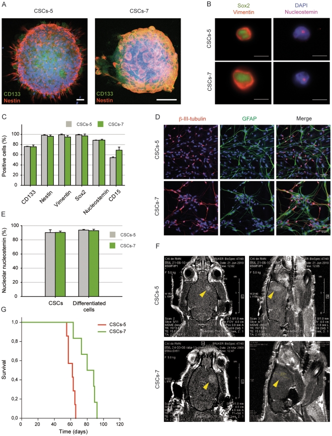
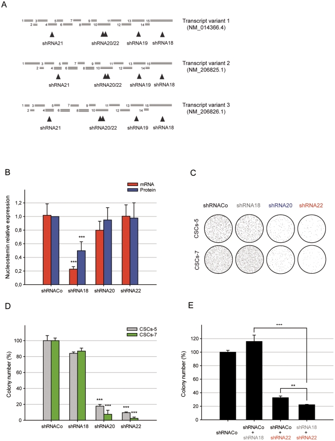
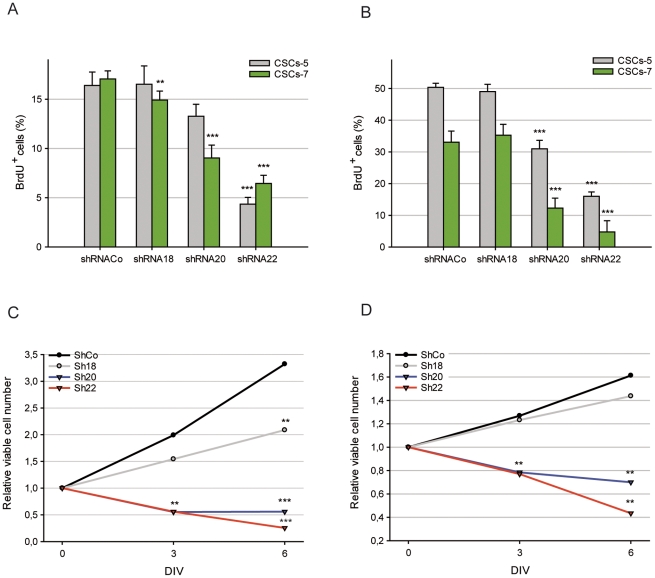
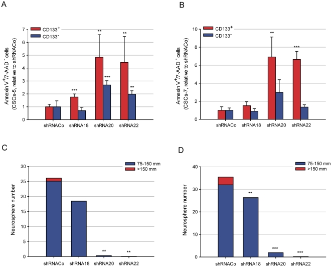
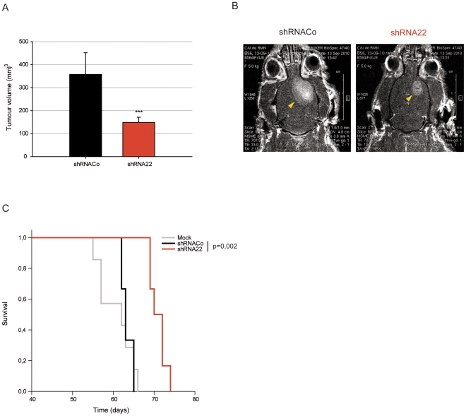
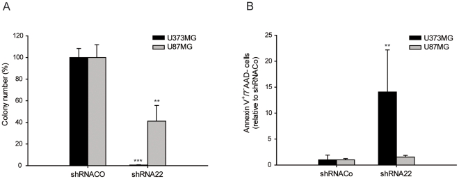
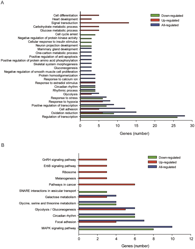
Glioblastomas (GBM) may contain a variable proportion of active cancer stem cells (CSCs) capable of self-renewal, of aggregating into CD133(+) neurospheres, and to develop intracranial tumors that phenocopy the original ones. We hypothesized that nucleostemin may contribute to cancer stem cell biology as these cells share characteristics with normal stem cells. Here we report that nucleostemin is expressed in GBM-CSCs isolated from patient samples, and that its expression, conversely to what it has been described for ordinary stem cells, does not disappear when cells are differentiated. The significance of nucleostemin expression in CSCs was addressed by targeting the corresponding mRNA using lentivirally transduced short hairpin RNA (shRNA). In doing so, we found an off-target nucleostemin RNAi (shRNA22) that abolishes proliferation and induces apoptosis in GBM-CSCs. Furthermore, in the presence of shRNA22, GBM-CSCs failed to form neurospheres in vitro or grow on soft agar. When these cells are xenotransplanted into the brains of nude rats, tumor development is significantly delayed. Attempts were made to identify the primary target/s of shRNA22, suggesting a transcription factor involved in one of the MAP-kinases signaling-pathways or multiple targets. The use of this shRNA may contribute to develop new therapeutic approaches for this incurable type of brain tumor.


Conflict of interest statement

Competing Interests: The authors have declared that no competing interests exist.

Figures

Figure 1
Figure 1. CSCs-5 and CSCs-7 characterization.
A. CSCs-5 and CSCs-7 neurospheres expressing CD133 (green) and nestin (red). Scalebar: 50 µm. B. CSCs-5 and CSCs-7 cells showing the expression and cellular localization of Sox2 (green), vimentin (red) and nucleostemin (purple). Scalebar: 10 µm, and quantitative plot (C) of the stem cell markers CD133, nestin, vimentin, Sox2, nucleostemin and CD15 in CSCs-5 (gray) and CSCs-7 (green). D. Neuron (β-III-tubulin, pink-red) and astrocytic (GFAP, green) differentiation capacity of CSCs-5 and CSCs-7. Scalebar: 50 µm. E. Nucleolar nucleostemin expression in stem or differentiatiated CSCs-5 (gray) and CSCs-7 (green) cells. F. Magnetic resonance imaging of in vivo tumors developed from CSCs-5 and CSCs-7 (yellow arrowheads point to the tumor), and Kaplan-Meier survival analysis (G) of immunodeficent rats, after CSCs-5 (red) or CSC-7 (green) orthotopic xenografts.
Figure 2
Figure 2. Nucleostemin-directed shRNAs effect in CSCs.
A. Schematic representation of the 3 nucleostemin transcript variants and exons. Arrowheads point to each designed shRNA. B. Nucleostemin mRNA (red) and protein (blue) quantification in CSCs-5 treated with shRNACo, shRNA18, shRNA20 and shRNA22. C. shRNAs effect on CSCs-5 and CSCs-7 soft agar colony-forming ability, and its quantitative analysis (D). E. Soft agar colony counts in double-infections to determine the nucleostemin-specificity of shRNA22. ** p≤0.05; *** p≤0.001.
Figure 3
Figure 3. shRNAs effect in the proliferation kinetic of CSCs.
A. Number of phase-S or total-cycling (B) CSCs-5 (grey) and CSCs-7 (green) cells treated with the different shRNAs. C. Number of viable cells on DIV 0, 3 and 6 in CSCs-5 and CSCs-7 (D) treated with the different shRNAs. ** p≤0.05; *** p≤0.001.
Figure 4
Figure 4. shRNAs effect in the viability of CSCs.
A. Apoptosis induced by shRNAs in CSCs-5 and CSCs-7 (B) CD133+ (red) or CD133 (blue) populations. C. Neurosphere forming-ability of CSCs-5 and CSCs-7 (D) cells, treated with the different shRNAs. ** p≤0.05; *** p≤0.001.
Figure 5
Figure 5. shRNAs effect in the in vivo tumor-development of the CSCs-5.
A. Quantification of the volumes of tumors induced with CSCs-5 cells treated with shRNACo (black) and shRNA22 (red). B. Magnetic resonance imaging of representative examples of tumors after orthotopic xenografts in nude rat-brains of CSCs-5 cells carrying shRNACo or shRNA22, and Kaplan-Meier survival plot (C) of the immunodeficent rats inoculated with CSCs-5 (mock, gray), cells carrying shRNACo (black) and shRNA22 (red). *** p≤0.001.
Figure 6
Figure 6. shRNAs effect in U373MG and U87MG cell lines.
A. shRNAs effect on U373MG (black) and U87MG (gray) soft agar colony-forming ability. B. shRNAs-induced apoptosis in U373MG (black) and U87MG (gray). ** p≤0.05; *** p≤0.001.
Figure 7
Figure 7. Gene expression profiling in the presence and absence of shRNA22.
A. Significative down- (green), up- (red) and all-regulated (blue) genes grouped by function of biological processes, or by belonging to signaling pathways (B) induced by shRNA22 in CSCs-5 cells.

Similar articles

Cited by

References

    1. Maher EA, Furnari FB, Bachoo RM, Rowitch DH, Louis DN, et al. Malignant glioma: genetics and biology of a grave matter. Genes Dev. 2001;15:1311–1333. - PubMed
    1. Stupp R, Mason WP, van den Bent MJ, Weller M, Fisher B, et al. Radiotherapy plus concomitant and adjuvant temozolomide for glioblastoma. N Engl J Med. 2005;352:987–996. - PubMed
    1. Clarke MF, Dick JE, Dirks PB, Eaves CJ, Jamieson CH, et al. Cancer stem cells–perspectives on current status and future directions: AACR Workshop on cancer stem cells. Cancer Res. 2006;66:9339–9344. - PubMed
    1. Lapidot T, Sirard C, Vormoor J, Murdoch B, Hoang T, et al. A cell initiating human acute myeloid leukaemia after transplantation into SCID mice. Nature. 1994;367:645–648. - PubMed
    1. Al-Hajj M, Wicha MS, Benito-Hernandez A, Morrison SJ, Clarke MF. Prospective identification of tumorigenic breast cancer cells. Proc Natl Acad Sci U S A. 2003;100:3983–3988. - PMC - PubMed

Publication types

MeSH terms