

Convergent evidence from multimodal imaging reveals amygdala abnormalities in schizophrenic patients and their first-degree relatives
- PMID: 22174900
- PMCID: PMC3234284
- DOI: 10.1371/journal.pone.0028794
Convergent evidence from multimodal imaging reveals amygdala abnormalities in schizophrenic patients and their first-degree relatives
Abstract
Background: Shared neuropathological features between schizophrenic patients and their first-degree relatives have potential as indicators of genetic vulnerability to schizophrenia. We sought to explore genetic influences on brain morphology and function in schizophrenic patients and their relatives.
Methods: Using a multimodal imaging strategy, we studied 33 schizophrenic patients, 55 of their unaffected parents, 30 healthy controls for patients, and 29 healthy controls for parents with voxel-based morphometry of structural MRI scans and functional connectivity analysis of resting-state functional MRI data.
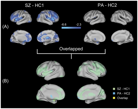
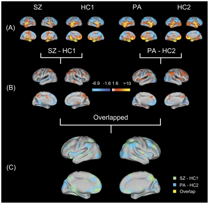
Results: Schizophrenic patients showed widespread gray matter reductions in the bilateral frontal cortices, bilateral insulae, bilateral occipital cortices, left amygdala and right thalamus, whereas their parents showed more localized reductions in the left amygdala, left thalamus and right orbitofrontal cortex. Patients and their parents shared gray matter loss in the left amygdala. Further investigation of the resting-state functional connectivity of the amygdala in the patients showed abnormal functional connectivity with the bilateral orbitofrontal cortices, bilateral precunei, bilateral dorsolateral frontal cortices and right insula. Their parents showed slightly less, but similar changes in the pattern in the amygdala connectivity. Co-occurrences of abnormal connectivity of the left amygdala with the left orbitofrontal cortex, right dorsolateral frontal cortex and right precuneus were observed in schizophrenic patients and their parents.
Conclusions: Our findings suggest a potential genetic influence on structural and functional abnormalities of the amygdala in schizophrenia. Such information could help future efforts to identify the endophenotypes that characterize the complex disorder of schizophrenia.
Conflict of interest statement
Figures
Similar articles
-
Similar and different gray matter deficits in schizophrenia patients and their unaffected biological relatives.Front Psychiatry. 2013 Nov 21;4:150. doi: 10.3389/fpsyt.2013.00150. Front Psychiatry. 2013. PMID: 24319433 Free PMC article. Review.
-
The Amygdala in Schizophrenia and Bipolar Disorder: A Synthesis of Structural MRI, Diffusion Tensor Imaging, and Resting-State Functional Connectivity Findings.Harv Rev Psychiatry. 2019 May/Jun;27(3):150-164. doi: 10.1097/HRP.0000000000000207. Harv Rev Psychiatry. 2019. PMID: 31082993 Review.
-
Neuroanatomical circuitry associated with exploratory eye movement in schizophrenia: a voxel-based morphometric study.PLoS One. 2011;6(10):e25805. doi: 10.1371/journal.pone.0025805. Epub 2011 Oct 3. PLoS One. 2011. PMID: 21991357 Free PMC article.
-
Association between functional and structural connectivity of the corticostriatal network in people with schizophrenia and unaffected first-degree relatives.J Psychiatry Neurosci. 2020 Nov 1;45(6):395-405. doi: 10.1503/jpn.190015. J Psychiatry Neurosci. 2020. PMID: 32436671 Free PMC article.
-
Impairments of thalamic resting-state functional connectivity in patients with chronic tinnitus.Eur J Radiol. 2015 Jul;84(7):1277-84. doi: 10.1016/j.ejrad.2015.04.006. Epub 2015 Apr 20. Eur J Radiol. 2015. PMID: 25935516
Cited by
-
Amygdala fMRI-A Critical Appraisal of the Extant Literature.Neurosci Insights. 2024 Aug 13;19:26331055241270591. doi: 10.1177/26331055241270591. eCollection 2024. Neurosci Insights. 2024. PMID: 39148643 Free PMC article. Review.
-
Similar and different gray matter deficits in schizophrenia patients and their unaffected biological relatives.Front Psychiatry. 2013 Nov 21;4:150. doi: 10.3389/fpsyt.2013.00150. Front Psychiatry. 2013. PMID: 24319433 Free PMC article. Review.
-
Disruptions in the left frontoparietal network underlie resting state endophenotypic markers in schizophrenia.Hum Brain Mapp. 2017 Apr;38(4):1741-1750. doi: 10.1002/hbm.23477. Epub 2016 Dec 23. Hum Brain Mapp. 2017. PMID: 28009080 Free PMC article.
-
Amygdala connectivity differs among chronic, early course, and individuals at risk for developing schizophrenia.Schizophr Bull. 2014 Sep;40(5):1105-16. doi: 10.1093/schbul/sbt165. Epub 2013 Dec 22. Schizophr Bull. 2014. PMID: 24366718 Free PMC article.
-
Amygdala signal abnormality and cognitive impairment in drug-naïve schizophrenia.BMC Psychiatry. 2023 Apr 5;23(1):231. doi: 10.1186/s12888-023-04728-6. BMC Psychiatry. 2023. PMID: 37020192 Free PMC article.
References
-
- Mueser KT, McGurk SR. Schizophrenia. Lancet. 2004;363:2063–2072. - PubMed
-
- Sawa A, Snyder SH. Schizophrenia: diverse approaches to a complex disease. Science. 2002;296:692–695. - PubMed
-
- Tsuang M. Schizophrenia: genes and environment. Biol Psychiatry. 2000;47:210–220. - PubMed
-
- McGuffin P, Owen MJ, Farmer AE. Genetic basis of schizophrenia. Lancet. 1995;346:678–682. - PubMed
Publication types
MeSH terms
LinkOut - more resources
Full Text Sources
Medical
