. 2011;6(12):e28915.
doi: 10.1371/journal.pone.0028915. Epub 2011 Dec 9.

Rosiglitazone inhibits transforming growth factor-β1 mediated fibrogenesis in ADPKD cyst-lining epithelial cells

Affiliations

Rosiglitazone inhibits transforming growth factor-β1 mediated fibrogenesis in ADPKD cyst-lining epithelial cells

Yawei Liu et al. PLoS One. 2011.

Abstract

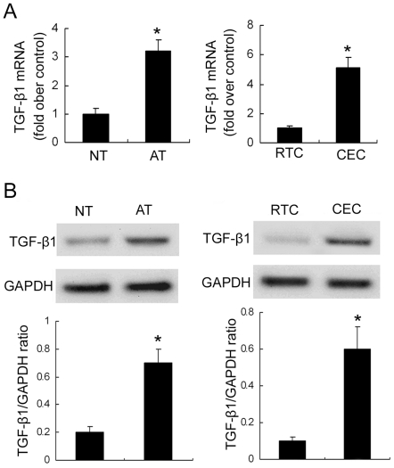
Background: Interstitial fibrosis plays an important role in progressive renal dysfunction in autosomal dominant polycystic kidney disease (ADPKD). In our previous studies, we confirmed that PPAR-γ agonist, rosiglitazone could protect renal function and prolong the survival of a slowly progressive ADPKD animal model by reducing renal fibrosis. However, the mechanism remains unknown.

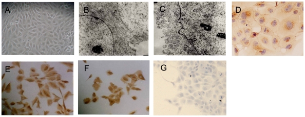
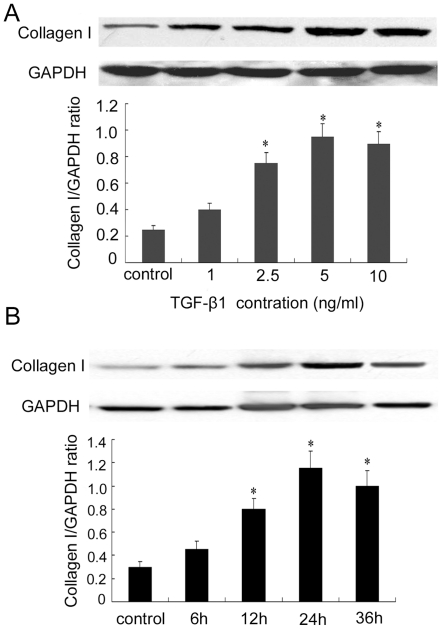
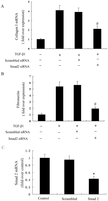
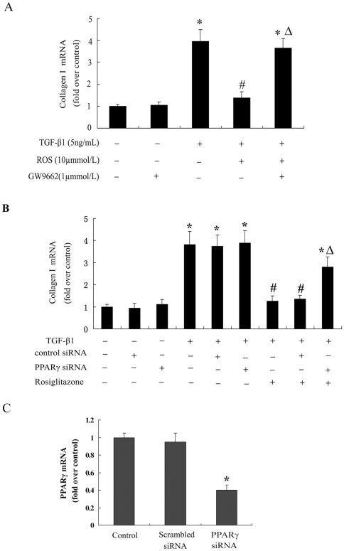
Methods: Primary culture epithelial cells pretreated with TGF-β1 were incubated with rosiglitazone. Extracellular matrix proteins were detected using real-time PCR and Western blotting. MAPK and Smad2 phosphorylation were measured with western blot. ERK1/2 pathway and P38 pathway were inhibited with the specific inhibitors PD98059 and SB203580. The Smad2 pathway was blocked with the siRNA. To address whether PPAR-γ agonist-mediated inhibition of TGF-β1-induced collagen type I expression was mediated through a PPAR-γ dependent mechanism, genetic and pharmaceutical approaches were used to block the activity of endogenous PPARγ.

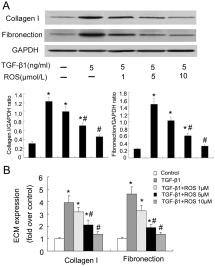
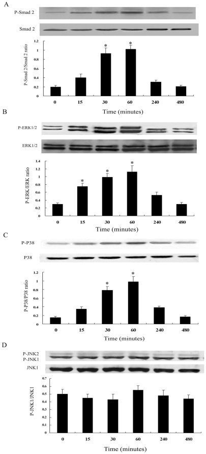
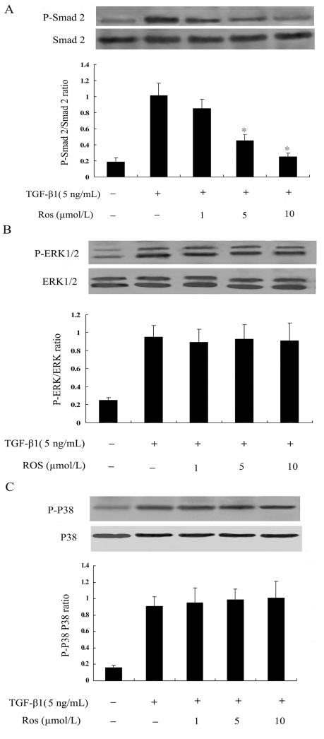
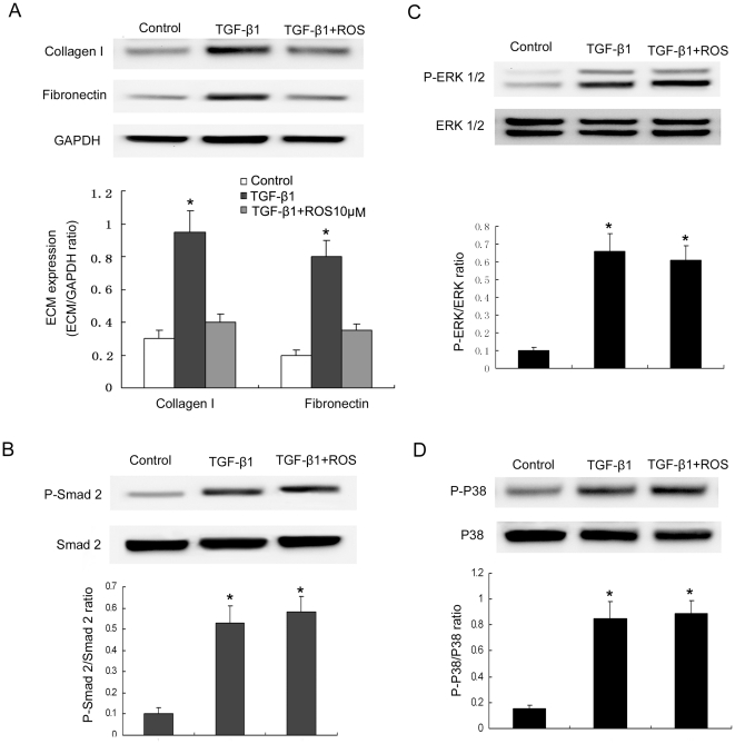
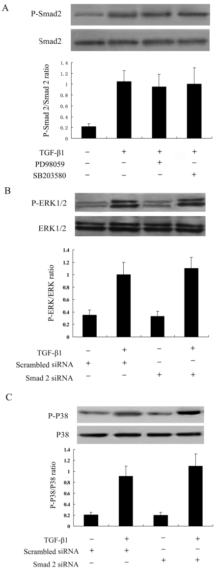
Results: TGF-β1-stimulated collagen type I and fibronectin expression of ADPKD cyst-lining epithelia were inhibited by rosiglitazone in a dosage-dependent manner. Smad2, ERK1/2 and P38 pathways were activated in response to TGF-β1; however, TGF-β1 had little effect on JNK pathway. Rosiglitazone suppressed TGF-β1 induced Smad2 activation, while ERK1/2 and P38MAPK signals remained unaffected. Rosiglitazone could also attenuate TGF-β1-stimulated collagen type I and fibronectin expression in primary renal tubular epithelial cells, but had no effect on TGF-β1-induced activation of Smad2, ERK1/2 and P38 pathways. There was no crosstalk between the Smad2 and MAPK pathways in ADPKD cyst-lining epithelial cells. These inhibitory effects of rosiglitazone were reversed by the PPARγ specific antagonist GW9662 and PPARγ siRNA.

Conclusion: ADPKD cyst-lining epithelial cells participate in TGF-β1 mediated fibrogenesis. Rosiglitazone could suppress TGF-β1-induced collagen type I and fibronectin expression in ADPKD cyst-lining epithelia through modulation of the Smad2 pathway. Our study may provide therapeutic basis for clinical applications of rosiglitazone in retarding the progression of ADPKD.


Conflict of interest statement

Competing Interests: The authors have declared that no competing interests exist.

Figures

Figure 1
Figure 1. Phase-contrast microscopic observation and identification of primary culture cyst-lining epithelial cells.
(A) Confluent monolayer of cyst-lining epithelial cells (Magnification ×200). (B–C). Transmission electron micrographs of cyst-lining epithelial cells (Magnification ×9600). B and C respectively showing the presence of adhesion plaque at tight junctions of cell-cell contact and microvilli-like coating. (D–G). Immunocytochemistry of E-cadherin (D) (Magnification ×400), cytokeratin (E) (Magnification ×200), vimentin (F) (Magnification ×200) and α-SMA (G) (Magnification ×200) in cyst-derived cells.
Figure 2
Figure 2. Expression of TGF-β1 in human normal kidney tissues, ADPKD kidney tissues, RTC and CEC.
(A) QRT-PCR analysis of TGF-β1 mRNA expression. (B) Immunoblotting analysis of TGF-β1 protein expression. Top panels were representative Western blot images. Bottom panels were the summary data from three independent experiments. *P<0.05 vs. control.
Figure 3
Figure 3. TGF-β1 induced collagen type I expression in a concentration- and time-dependent manner in ADPKD cyst-lining epithelial cells.
(A) TGF-β1 treatment (1–10 ng/mL, 24 h). (B) TGF-β1 treatment (5 ng/mL, 6–36 h). Top panels were representative Western blot images. Bottom panels were the summary data from three independent experiments. *P<0.05 vs. control.
Figure 4
Figure 4. Rosiglitazone inhibited TGF-β1-induced collagen type I and fibronectin protein and mRNA synthesis in ADPKD cyst-lining epithelial cells in a concentration-dependent fashion.
Cells were pretreated with rosiglitazone for 1 h, and then incubated with TGF-β1 for 24 h. A. collagen type I and fibronectin protein in TGF-β1-stimulated ADPKD cyst-lining epithelial cells treated with rosiglitazone. B. collagen type I and fibronectin mRNA in TGF-β1-stimulated ADPKD cyst-lining epithelial cells treated with rosiglitazone. All results were representative of three independent experiments with similar results. *P<0.05 vs. control; #P<0.05 vs. TGF-β1.
Figure 5
Figure 5. Time course of Smad2 (A), ERK1/2 (B), p38MAPK (C) and JNK (D) activation by TGF-β1 in ADPKD cyst-lining epithelial cells.
Cells were treated with TGF-β1 (5 ng/ml) for the indicated time periods. Kinase activation was determined by western blot analysis using phosphospecific antibodies. As controls, the protein levels of Smad2, ERK1/2, p38MAPK and JNK were determined using corresponding non-phosphorylated form antibodies. Results of densitometric analysis, expressed as a ratio between phospho- and non-phospho-antibody, from three independent experiments were shown. *P<0.05 vs. control.
Figure 6
Figure 6. Evaluation of inhibitory effect of rosiglitazone on TGF-β1 induced Smad2 (A), ERK1/2 (B) and p38MAPK(C) activation in ADPKD cyst-lining epithelial cells.
Cells were pretreated with rosiglitazone for 1 h, and then incubated with rosiglitazone in the presence or absence of TGF-β1 (5 ng/mL) for another 1h. * P<0.05 vs. TGF-β1 alone.
Figure 7
Figure 7. Effect of Smad2 siRNA on TGF-β1 induced collagen type I and fibronectin mRNA expression.
Smad2 was inhibited using the Smad2 siRNA method. Cells were transfected with Smad2 siRNA for 48 h, followed by treatment with TGF-β1 for 24 h. (A) Smad2 siRNA significantly reduced collagen type I synthesis in TGF-β1-stimulated cells. (B) Smad2 siRNA significantly reduced fibronectin synthesis in TGF-β1-stimulated cells. (C) Smad2 mRNA was decreased to 37.8% using real-time RT–PCR in Smad2 siRNA-transfected ADPKD cyst-lining epithelial cells. The results were representative of three independent experiments. *P<0.05 vs. control, # P<0.05 vs. TGF-β1 alone.
Figure 8
Figure 8. The role of rosiglitazone on TGF-β1-induced primary renal tubular epithelial cells.
Collagen type I and fibronectin expression in TGF-β1-stimulated primary renal tubular epithelial cells treated with rosiglitazone (A). Cells were pretreated with rosiglitazone (10 µmol/L) for 1 h, and then incubated with TGF-β1 for 24 h. Evaluation of inhibitory effect of rosiglitazone on TGF-β1-induced Smad2 (B), ERK1/2 (C) and p38MAPK (D) activation in primary renal tubular epithelial cells. Cells were pretreated with rosiglitazone for 1 h, and then incubated with rosiglitazone in the presence or absence of TGF-β1 (5 ng/mL) for another 1 h. *P<0.05 vs. control.
Figure 9
Figure 9. Crosstalk between Smad2 and MAPK signals in TGF-β1-stimulated ADPKD cyst-lining epithelial cells.
(A) Smad2 activation was not affected by inhibition of ERK or P38 MAPK pathways. ADPKD cyst-lining epithelial cells pre-treated with PD98059 (25 µM) or SB203580 (10 µM) for 1 h were stimulated with TGF-β1 for another 1 h. (B) ERK activation was not affected by inhibition of the Smad2 signal. Smad2 was inhibited using Smad2 siRNA method. (C) P38 activation was not affected by inhibition of the Smad2 signal. The results were representative of three independent experiments.
Figure 10
Figure 10. Rosiglitazone inhibited TGF-β1-induced collagen type I expression in ADPKD cyst-lining epithelial cells through PPARγ.
(A) Cells were incubated with GW9662 (1 µmol/L) and cultured for 48 h. Then cells were pretreated with 10 µmol/L rosiglitazone for 1 h and incubated with TGF-β1 (5 ng/mL) for 24 h. (B) Cells were transfected with a PPARγ siRNA for 48 h, then pretreated with 10 µmol/L rosiglitazone for 1 h and incubated with TGF-β1(5 ng/mL) for 24 h. (C) PPARγ mRNA was decreased to 36% using real-time RT–PCR in PPARγ siRNA-transfected ADPKD cyst-lining epithelial cells. The results were representative of three independent experiments. *P<0.05 vs. control; #P<0.05 vs. TGF-β1 alone Δ P<0.05 vs. rosiglitazone + TGF-β1.

Similar articles

Cited by

References

    1. Torres VE, Harris PC, Pirson Y. Autosomal dominant polycystic kidney disease. Lancet. 2007;369:1287–1301. - PubMed
    1. Harris PC, Torres VE. Polycystic kidney disease. Annu Rev Med. 2009;60:321–337. - PMC - PubMed
    1. Torres VE, Harris PC. Mechanisms of Disease: autosomal dominant and recessive polycystic kidney diseases. Nat Clin Pract Nephrol 2: 40-55; quiz. 2006;55 - PubMed
    1. Lieske JC, Toback FG. Autosomal dominant polycystic kidney disease. J Am Soc Nephrol. 1993;3:1442–1450. - PubMed
    1. Hassane S, Leonhard WN, van der Wal A, Hawinkels LJ, Lantinga-van Leeuwen IS, et al. Elevated TGFbeta-Smad signalling in experimental Pkd1 models and human patients with polycystic kidney disease. J Pathol. 2010;222:21–31. - PubMed

Publication types

MeSH terms