

One-year results of photodynamic therapy combined with intravitreal ranibizumab for exudative age-related macular degeneration
- PMID: 22174997
- PMCID: PMC3235818
- DOI: 10.1155/2012/154659
One-year results of photodynamic therapy combined with intravitreal ranibizumab for exudative age-related macular degeneration
Abstract
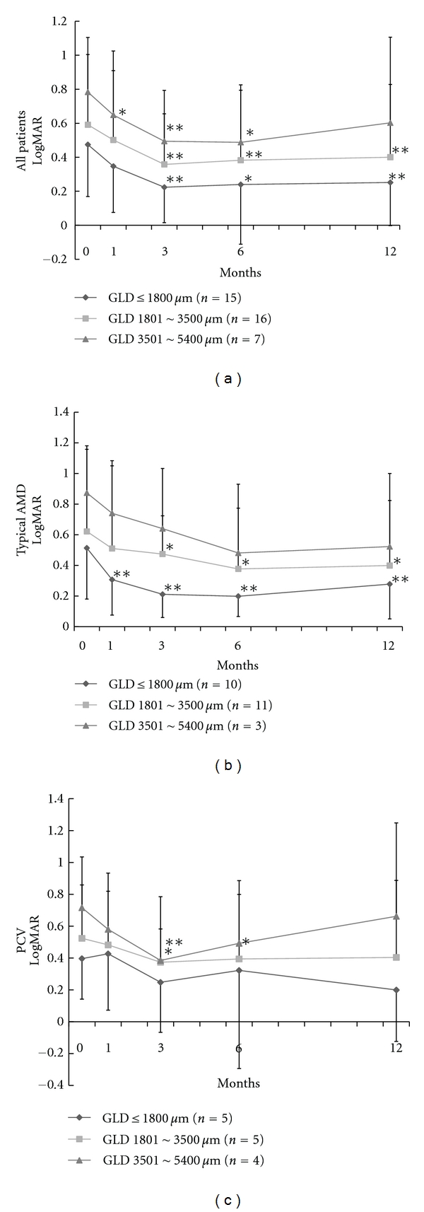
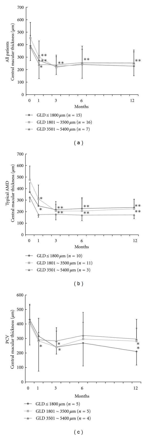
Purpose. To evaluate the effects of photodynamic therapy (PDT) combined with intravitreal injection of ranibizumab (IVR) for exudative age-related macular degeneration (AMD). Methods. Retrospective case series. Thirty eight eyes of 38 patients with exudative AMD underwent combined therapy consisting first of IVR, followed by PDT within a week and the second IVR at 1 month. All patients were followed up for more than 12 months. The best corrected visual acuity (BCVA) and central macular thickness (CMT) were examined. Results. The mean number of IVR and PDT sessions were 2.9 ± 1.3 and 1.1 ± 0.3, respectively. The mean BCVA and CMT were significantly improved to 0.38 logMAR units (P < 0.01) and 240 μm (P < 0.01) at 12 months, respectively. Thirty-six of 38 eyes (94.8%) improved or maintained BCVA at 12 months. Conclusion. PDT combined with IVR for exudative AMD was effective at improving visual acuity and CMT with a low recurrence rate for 12 months.
Figures
Similar articles
-
Effectiveness of intravitreal ranibizumab in exudative age-related macular degeneration (AMD): comparison between typical neovascular AMD and polypoidal choroidal vasculopathy over a 1 year follow-up.BMC Ophthalmol. 2013 Apr 4;13:10. doi: 10.1186/1471-2415-13-10. BMC Ophthalmol. 2013. PMID: 23557322 Free PMC article.
-
Intravitreal Ranibizumab Versus Intravitreal Ranibizumab Combined with Posterior Subtenon Triamcinolone Acetonide in Diabetic Macular Edema.Beyoglu Eye J. 2021 Sep 27;6(3):229-235. doi: 10.14744/bej.2021.53315. eCollection 2021. Beyoglu Eye J. 2021. PMID: 35005521 Free PMC article.
-
Effect of switching therapy to pegaptanib in eyes with the persistent cases of exudative age-related macular degeneration.Medicine (Baltimore). 2014 Oct;93(18):e116. doi: 10.1097/MD.0000000000000116. Medicine (Baltimore). 2014. PMID: 25319441 Free PMC article.
-
Comparison of one-year results of photodynamic therapy combined with ranibizumab or aflibercept for treating polypoidal choroidal vasculopathy.PLoS One. 2020 Jun 24;15(6):e0235213. doi: 10.1371/journal.pone.0235213. eCollection 2020. PLoS One. 2020. PMID: 32579608 Free PMC article.
-
Intravitreal Ranibizumab Alone or in Combination with Calcium Dobesilate for the Treatment of Diabetic Macular Edema in Nonproliferative Diabetic Retinopathy Patients: 12-Month Outcomes of a Retrospective Study.Int J Clin Pract. 2022 Oct 20;2022:6725225. doi: 10.1155/2022/6725225. eCollection 2022. Int J Clin Pract. 2022. PMID: 36340967 Free PMC article. Review.
Cited by
-
Efficacy of ranibizumab combined with photodynamic therapy on wet age-related macular degeneration.Exp Ther Med. 2020 Jun;19(6):3691-3697. doi: 10.3892/etm.2020.8641. Epub 2020 Apr 2. Exp Ther Med. 2020. PMID: 32346433 Free PMC article.
References
-
- Zarbin MA. Current concepts in the pathogenesis of age-related macular degeneration. Archives of Ophthalmology. 2004;122(4):598–614. - PubMed
-
- Tzekov R, Lin T, Zhang K-M, et al. Ocular changes after photodynamic therapy. Investigative Ophthalmology and Visual Science. 2006;47(1):377–385. - PubMed
-
- Lai TYY, Chan WM, Lam DSC. Transient reduction in retinal function revealed by multifocal electroretinogram after photodynamic therapy. American Journal of Ophthalmology. 2004;137(5):826–833. - PubMed
-
- Kvanta A, Algvere PV, Berglin L, Seregard S. Subfoveal fibrovascular membranes in age-related macular degeneration express vascular endothelial growth factor. Investigative Ophthalmology and Visual Science. 1996;37(9):1929–1934. - PubMed
-
- Boyer DS, Antoszyk AN, Awh CC, Bhisitkul RB, Shapiro H, Acharya NR. Subgroup analysis of the MARINA study of ranibizumab in neovascular age-related macular degeneration. Ophthalmology. 2007;114(2):246–252. - PubMed
LinkOut - more resources
Full Text Sources
