

Reactive oxygen species in skeletal muscle signaling
- PMID: 22175016
- PMCID: PMC3235811
- DOI: 10.1155/2012/982794
Reactive oxygen species in skeletal muscle signaling
Abstract
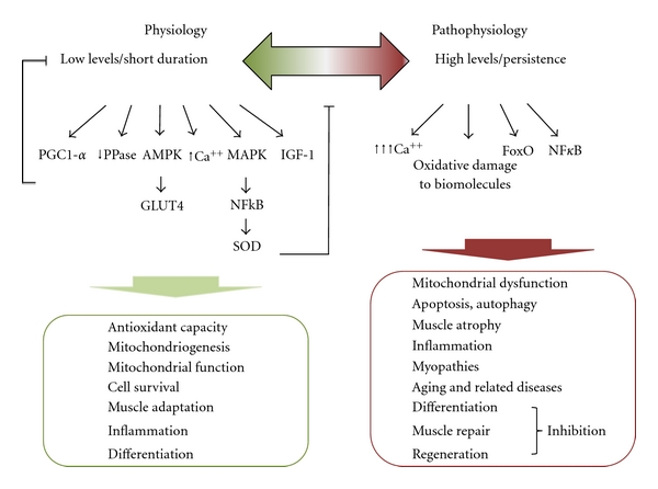
Generation of reactive oxygen species (ROS) is a ubiquitous phenomenon in eukaryotic cells' life. Up to the 1990s of the past century, ROS have been solely considered as toxic species resulting in oxidative stress, pathogenesis and aging. However, there is now clear evidence that ROS are not merely toxic species but also-within certain concentrations-useful signaling molecules regulating physiological processes. During intense skeletal muscle contractile activity myotubes' mitochondria generate high ROS flows: this renders skeletal muscle a tissue where ROS hold a particular relevance. According to their hormetic nature, in muscles ROS may trigger different signaling pathways leading to diverging responses, from adaptation to cell death. Whether a "positive" or "negative" response will prevail depends on many variables such as, among others, the site of ROS production, the persistence of ROS flow or target cells' antioxidant status. In this light, a specific threshold of physiological ROS concentrations above which ROS exert negative, toxic effects is hard to determine, and the concept of "physiologically compatible" levels of ROS would better fit with such a dynamic scenario. In this review these concepts will be discussed along with the most relevant signaling pathways triggered and/or affected by ROS in skeletal muscle.
Figures
References
-
- Murphy ME, Kehrer JP. Activities of antioxidant enzymes in muscle, liver and lung of chickens with inherited muscular dystrophy. Biochemical and Biophysical Research Communications. 1986;134(2):550–556. - PubMed
-
- Tidball JG. Inflammatory processes in muscle injury and repair. American Journal of Physiology—Regulatory Integrative and Comparative Physiology. 2005;288(2):R345–R353. - PubMed
-
- Adhihetty PJ, Irrcher I, Joseph AM, Ljubicic V, Hood DA. Plasticity of skeletal muscle mitochondria in response to contractile activity. Experimental Physiology. 2003;88(1):99–107. - PubMed
-
- Rochard P, Rodier A, Casas F, et al. Mitochondrial activity is involved in the regulation of myoblast differentiation through myogenin expression and activity of myogenic factors. Journal of Biological Chemistry. 2000;275(4):2733–2744. - PubMed
-
- Sestili P, Barbieri E, Martinelli C, et al. Creatine supplementation prevents the inhibition of myogenic differentiation in oxidatively injured C2C12 murine myoblasts. Molecular Nutrition and Food Research. 2009;53(9):1187–1204. - PubMed
LinkOut - more resources
Full Text Sources
Other Literature Sources
Medical
Miscellaneous
