. 2011:2011:296151.
doi: 10.1155/2011/296151. Epub 2011 Nov 16.

The Trojan Horse Liposome Technology for Nonviral Gene Transfer across the Blood-Brain Barrier

Affiliations

The Trojan Horse Liposome Technology for Nonviral Gene Transfer across the Blood-Brain Barrier

Ruben J Boado et al. J Drug Deliv. 2011.

Abstract

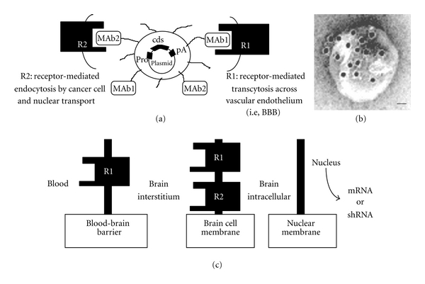
The application of blood-borne gene therapy protocols to the brain is limited by the presence of the blood-brain barrier (BBB). Viruses have been extensively used as gene delivery systems. However, their efficacy in brain is limited by the lack of transport across the BBB following intravenous (IV) administration. Recent progress in the "Trojan Horse Liposome" (THL) technology applied to transvascular non-viral gene therapy of the brain presents a promising solution to the trans-vascular brain gene delivery problem. THLs are comprised of immunoliposomes carrying nonviral gene expression plasmids. The tissue target specificity of the THL is provided by peptidomimetic monoclonal antibody (MAb) component of the THL, which binds to specific endogenous receptors located on both the BBB and on brain cellular membranes, for example, insulin receptor and transferrin receptor. These MAbs mediate (a) receptor-mediated transcytosis of the THL complex through the BBB, (b) endocytosis into brain cells and (c) transport to the brain cell nuclear compartment. The expression of the transgene in brain may be restricted using tissue/cell specific gene promoters. This manuscript presents an overview on the THL transport technology applied to brain disorders, including lysosomal storage disorders and Parkinson's disease.


Figures

Figure 1
Figure 1
Engineering of Trojan horse liposomes (THL). (a) A supercoiled plasmid DNA is encapsulated in the interior of the THL. The plasmid encodes for a coding sequence (cds), the expression of which is under the influence of a promoter (pro), that is, SV40, and a polyadenylation sequence (pA). The surface of the liposome contains several thousand strands of 2000 Da polyethylene glycol (PEG) to stabilize the complex in blood. Approximately 1-2% of the PEG strands are conjugated with a targeting receptor- (R-) specific monoclonal antibody (MAb) (Table 1), which triggers transport of the THL across biological barriers in vivo. THLs are engineered with a single type of MAb to target both the BBB and brain cells in the same species. In an experimental mouse model of a human brain tumor, the THL is engineered with both the 8D3 mouse transferrin receptor (TfR) MAb (MAb1) to target the mouse BBB (i.e., R1) and the 8314 human IR MAb (MAb2) to target the human tumor cells (i.e., R2). Thus, the THL is transported through the mouse BBB via receptor-mediated transcytosis on the mouse TfR, and then through the intracranial human glioma cell membrane via endocytosis on the human insulin receptor. (b) Transmission electron microscopy of a THL. Mouse IgG molecules tethered to the tips of the PEG strands on the surface of the THL were detected with a conjugate of 10 nm gold and an antimouse secondary antibody. The position of the gold particles illustrates the relationship of the PEG-extended MAb and the liposome surface. Magnification bar = 20 nm. (c) The 3-barrier model for gene therapy of the brain. Following intravenous injection, the THL carrying the transgene must traverse 3 barriers in series to be able to reach the nucleus for expression: (a) the blood-brain barrier (BBB), (b) the brain cell membrane (BCM), and (c) the nuclear membrane. THLs can be engineered with a single type of MAb to target the same receptor in both the BBB and BCM (R1) or with 2 different MAbs to target different receptors at the BBB and the BCM, for example, R1 and R2, respectively. From [4].
Figure 2
Figure 2
In vivo gene expression of a β-galactosidase reporter gene following systemic administration of THLs. (Top panels) β-galactoside histochemistry was performed on mouse brain and spleen removed 2 days after an IV injection of THLs carrying a β-galactosidase plasmid driven by either the SV40 promoter (SV40-lacZ-THL) (left panels) or Gfap promoter (Gfap-lacZ-THL) (right panels). THLs were targeted with the 8D3 antimouse TfRMAb. (Bottom panels) β-Galactosidase histochemistry of Rhesus monkey brain removed from either a monkey injected with THLs targeted with the HIRMAb ((a), (c), (d), (e), and (f)) or a control uninjected primate (b). The β-galactosidase expression plasmid is driven by the SV40 promoter. (a) shows a full coronal section of the primate forebrain. (c) shows half-coronal sections through the primate cerebrum and a full coronal section through the cerebellum; the sections from top to bottom are taken from the rostral to caudal parts of brain. (d, e, and f) are light micrographs of choroid plexus, occipital cortex, and cerebellum, respectively. All specimens are β-galactosidase histochemistry without counterstaining. The magnification in (a) and (b) is the same and the magnification bar in (a) is 3 mm; the magnification bar in (c) is 8 mm; the magnification bars in (d)–(f) are 155 μm. Top panels are from [21]. Bottom panels are from [34].
Figure 3
Figure 3
Enzyme replacement therapy with THLs in a mouse model of type VII mucopolysaccharidosis. (a) GUSB enzyme activity in GUSB null (−) fibroblasts and in fibroblasts obtained from wild type (+) mice. Fibroblasts were treated either with saline or with TfRMAb-targeted THLs encapsulated with the pCMV-GUSB expression plasmid. Data are mean ± SE (n = 4), and statistical significance was determined with ANOVA and Dunnett's test. The difference in GUSB enzyme activity in the THL-treated cells is significantly different from the untreated cells from the GUSB null mice (P < 0.01). (b) GUSB enzyme activity in brain and five other organs of GUSB null mice removed at 48 h after single intravenous administration of either saline or 10 ug/mouse of pCMV-GUSB plasmid DNA encapsulated in TfRMAb-targeted THLs. Mean ± SE (n = 4-5 mice/group). The difference in GUSB enzyme activity in the THL-treated mice, as compared to the saline-treated mice, is significant (P < 0.0005), in all organs, except the heart. From [36].
Figure 4
Figure 4
Levels of TH in brain following TH-gene therapy in the 6-OHDA Parkinson's disease model. The TH immunocytochemistry was performed in rat brains removed 72 hours after a single intravenous injection of 10 μg per rat of clone 951 plasmid DNA encapsulated in THL targeted with either the TfRMAb (a, b, and c) or with the mouse IgG2a isotype control (d, e, and f). Coronal sections are shown for 3 different rats from each of the two treatment groups. The 6-hydroxydopamine was injected in the medial forebrain bundle of the right hemisphere, which corresponds to right side of the figure. Sections are not counterstained. The animals that received the TH gene therapy had a normalization of the brain TH levels as compared to the animals administered the nontargeted THLs, which showed complete lost of immunoreactive TH in the same region. From [30].
Figure 5
Figure 5
Enzyme replacement therapy in a Parkinson's disease model using THLs and TH genomic expression vectors. (a) Diagrams of four rat TH expression plasmids. The poly(A) transcription termination sequence is the SV40 3′-untranslated region (UTR) derived from the pGL2 promoter vector (Promega) for both clone 877 and prgTH4, whereas the poly(A) signal for prgTH2 and prgTH3 is derived from the rat TH gene. (b) Striatal TH enzyme activity on the side ipsilateral to the toxin lesion at 3, 6, and 10 days after a single injection of either saline or THLs carrying clone 877 alone, prgTH3 alone, or clone 877 + prgTH3 combined. Data are mean ± S.E. (n = 3–6 rats per point), and statistically significant differences were determined by Student's t-test. All plasmids were delivered to rat brain following the intravenous injection of TfRMAb-targeted THLs. The TH activity at 3 days following combination therapy is significantly greater than prgTH3 alone at 3 and 6 days after injection (P < 0.005) and is greater than clone 877 alone at 10 days after injection (P < 0.005). (c) Apomorphine-induced rotations at 3, 6, and 10 days after a single injection of either saline or THLs carrying clone 877 alone, prgTH3 alone, or clone 877 + prgTH3 combined. Data are mean ± S.E. (n = 3–6 rats per point). The rotation behavior at 3 days following combination therapy is significantly reduced as compared to prgTH3 alone at 3 days after injection (P < 0.005) and is significantly reduced as compared to clone 877 alone at 10 days after injection (P < 0.005). From [48].

Similar articles

Cited by

References

    1. Pardridge WM. Drug and gene delivery to the brain: the vascular route. Neuron. 2002;36(4):555–558. - PubMed
    1. Mok KW, Lam AM, Cullis PR. Stabilized plasmid-lipid particles: factors influencing plasmid entrapment and transfection properties. Biochimica et Biophysica Acta. 1999;1419(2):137–150. - PubMed
    1. Shi N, Pardridge WM. Noninvasive gene targeting to the brain. Proceedings of the National Academy of Sciences of the United States of America. 2000;97(13):7567–7572. - PMC - PubMed
    1. Boado RJ. Blood-brain barrier transport of non-viral gene and RNAi therapeutics. Pharmaceutical Research. 2007;24(9):1772–1787. - PubMed
    1. Byrnes AP, Rusby JE, Wood MJ, Charlton HM. Adenovirus gene transfer causes inflammation in the brain. Neuroscience. 1995;66(4):1015–1024. - PubMed

LinkOut - more resources