. 2012 May;24(5):789-97.
doi: 10.1111/j.1365-2826.2011.02272.x.

Interaction between P2X3 and oestrogen receptor (ER)α/ERβ in ATP-mediated calcium signalling in mice sensory neurones

Affiliations

Interaction between P2X3 and oestrogen receptor (ER)α/ERβ in ATP-mediated calcium signalling in mice sensory neurones

T Cho et al. J Neuroendocrinol. 2012 May.

Abstract

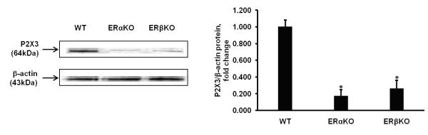
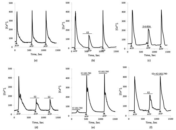
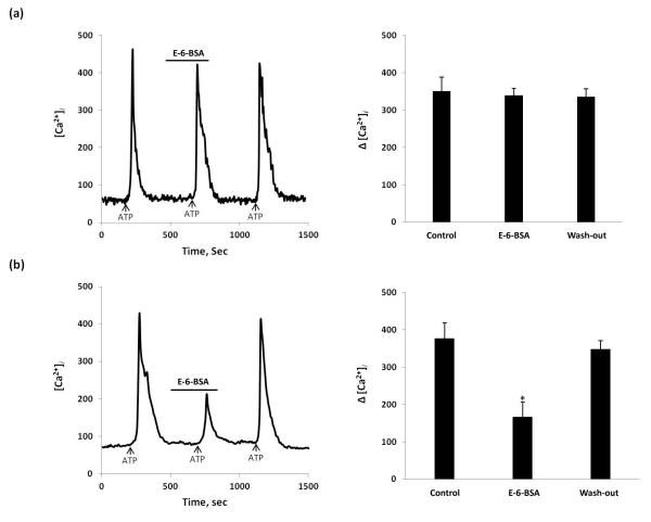
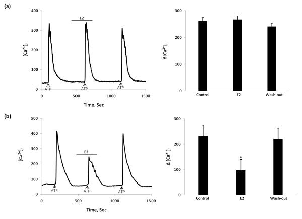
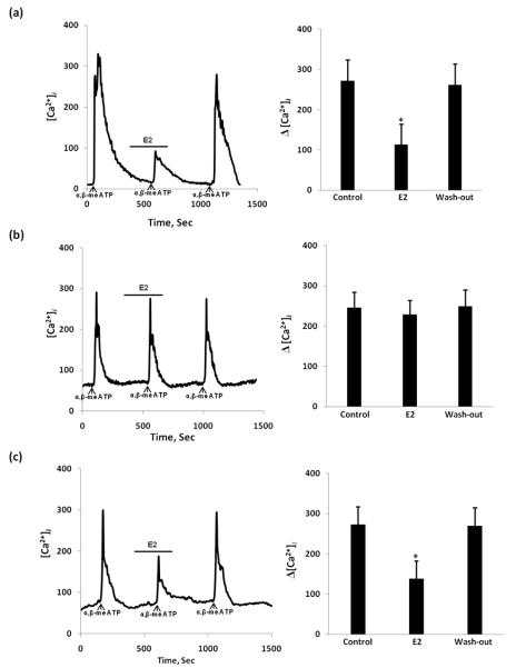
Emerging evidence supports a role of purinergic P2X3 receptors in modulating nociceptive signalling in sensory neurones. Previously, we showed that dorsal root ganglion (DRG) neurones (L1-S1) express both oestrogen receptor (ER)α and ERβ receptors. In the present study, we investigated the expression of P2X3 receptors and the effect of 17β-oestradiol (E(2)) on the ATP-induced [Ca(2+)](i) increase in DRG neurones collected from C57Bl/6J, ERα knockout (KO) and ERβKO mice. Our data showed a significant decrease for P2X3 in ERαKO (all levels) and ERβKO (mostly observed in L1, L2, L4 and L6). Furthermore, E(2) (100 nm) significantly attenuated the ATP (10 μm)-induced [Ca(2+)](i) in C57Bl/6J mice. ER antagonist ICI 182,780 (1 μm) blocked this attenuation. Homomeric P2X3 receptors are plentifully expressed in DRG neurones and contribute to nociceptive signals. α,β-Methylene (α,β-me) ATP, which is a specific agonist of P2X2/3 receptors, showed similar responses to the ATP-induced calcium increase in KO mice. A membrane-impermeable E-6-bovine serum albumin (1 μm) had the same effect as E(2) , suggesting action on the membrane. In DRG neurones from ERβKO and wild-type mice, E(2) attenuated the ATP/α,β-me ATP-induced [Ca(2+)](i) fluxes but, in DRG neurones from ERαKO mice, this hormone had no effect, suggesting that this attenuation depends on membrane-associated ERα receptors. Together, our data indicate an interaction between P2X3 and membrane-associated ERα in primary sensory neurones that may represent a novel mechanism to explain sex differences observed in the clinical presentation of visceral nociceptive syndromes.


Figures

Figure 1
Figure 1
Western blot analysis of DRG lysates shows reduced expression of P2X3 in both knock-out mice. Quantification of signals from Western blots shows statistically significant difference between the intensity of the bands from both knock-out DRG neurons when compared with wild type animals.
Figure 2
Figure 2
(a) Expression of P2X3 receptors in DRG neurons from Wt, ERαKO, and ERβKO in vivo using fluorescent microscopy. DRG sections (L2 level) were incubated in P2X3 primary antibodies. (b) Percentage distribution of labeled dorsal root ganglion neurons in ERαKO and ERβKO as well as wild type mice with P2X3 through L1-S1 levels. * indicate statistically significant difference from control, P<0.05.
Figure 3
Figure 3
17β-estradiol (E2) inhibits ATP-induced [Ca2+]i transients in wild type mice. (a) Typical indication of equal [Ca2+]i responses to repeated ATP (10 μM) stimulation (indicated by arrow) with 10 min interval under control condition. (b) Second ATP-induced [Ca2+]i response rapidly attenuated by E2 (100 nM) in dorsal root ganglion cells. (c) E2-BSA (1 μM) inhibited the ATP-induced [Ca2+]i transient. After wash-out with experimental medium, ATP response on [Ca2+]i returned to initial (control) amplitude of stimulation. (d) The effect of E2 does not desensitize upon repeated application of ATP. (e) Effect of estrogen receptor antagonist ICI 182,780 alone and application of ATP. (f) ICI 182,780 (1 μM) blocked the E2 attenuation of ATP-induced [Ca2+]i transients.
Figure 4
Figure 4
Summary of ATP-induced [Ca2+]i influxes in control, in the presence of E2, E-6-BSA, and ICI 182,780. E2 significantly decreased [Ca2+]i response to ATP whereas estrogen receptor antagonist ICI 182,780 blocked E2 effect. Values are expressed as mean ± SEM. * indicate statistically significant difference from control, P<0.05.
Figure 5
Figure 5
The effect of E-6-BSA on ATP-induced [Ca2+]i fluxes in ERαKO and ERβKO mice. (a) In ERαKO mouse, E-6-BSA added for 5 min didn’t inhibit ATP-induced [Ca2+]i flux; (b) In ERβKO mouse, Effect of E-6-BSA mimicked that observed in Wt mouse. Summary data represented on the right bar graphs. * Statistically significant difference from control, P<0.05.
Figure 6
Figure 6
The effect of E2 on ATP-induced [Ca2+]i transients in estrogen receptor-α knockout (ERαKO) and estrogen receptor-β knockout (ERβKO) mice. (a) In ERαKO mouse, E2 added for 5 min didn’t inhibit ATP-induced [Ca2+]i transient; (b) In ERβKO mouse E2 stimulation significantly attenuated the ATP-stimulated [Ca2+]i transient similar to that observed in Wt mouse. Summary data represented on the right bar graphs. *Statistically significant difference from control, P<0.05.
Figure 7
Figure 7
The effect of 17β-estradiol (E2) on α,β-meATP-induced [Ca2+]i transients in Wt, ERαKO, and ERβKO mice. (a) Wt mouse as a control, (b) ERαKO mouse, E2 added for 5min didn’t inhibit α,β-meATP-induced [Ca2+]i transient in control vs. after E2 treatment. (c) In ERβKO mouse, E2 stimulation significantly attenuated the α,β-meATP-stimulated [Ca2+]i transient similar to that observed in Wt mouse. Summary data represented on the right bar graphs. Values are expressed as mean±SEM. Δ[Ca2+]i were determined by subtracting the [Ca2+]i peak levels from the basal [Ca2+]i levels. *Statistically significant difference from control, p < 0.05.

References

    1. Chang L, Heitkemper MM. Gender differences in irritable bowel syndrome. Gastroenterology. 2002;123(5):1686–701. - PubMed
    1. Berkley KJ, Zalcman SS, Simon VR. Sex and gender differences in pain and inflammation: a rapidly maturing field. Am J Physiol Regul Integr Comp Physiol. 2006;291(2):R241–4. - PubMed
    1. Nadal A, Diaz M, Valverde MA. The estrogen trinity: membrane, cytosolic, and nuclear effects. News Physiol Sci. 2001;16:251–5. - PubMed
    1. Levin ER. Cellular functions of plasma membrane estrogen receptors. Steroids. 2002;67(6):471–5. - PubMed
    1. Razandi M, et al. Identification of a structural determinant necessary for the localization and function of estrogen receptor alpha at the plasma membrane. Mol Cell Biol. 2003;23(5):1633–46. - PMC - PubMed

Publication types

MeSH terms