. 2011 Dec 16:9:217.
doi: 10.1186/1479-5876-9-217.

Endogenous interleukin-10 constrains Th17 cells in patients with inflammatory bowel disease

Affiliations

Endogenous interleukin-10 constrains Th17 cells in patients with inflammatory bowel disease

Cailin M Wilke et al. J Transl Med. .

Abstract

Background: Th17 cells play a role in inflammation. Interleukin (IL)-10 is a potent anti-inflammatory cytokine. However, it is poorly understood whether and how endogenous IL-10 impacts the development of Th17 cells in human pathologies.

Materials and methods: We examined the relationship between IL-10 and Th17 cells in patients with Crohn's disease and in IL-10-deficient (IL-10-/-) mice. Th17 cells and dendritic cells (DCs) were defined by flow cytometry and evaluated by functional studies.

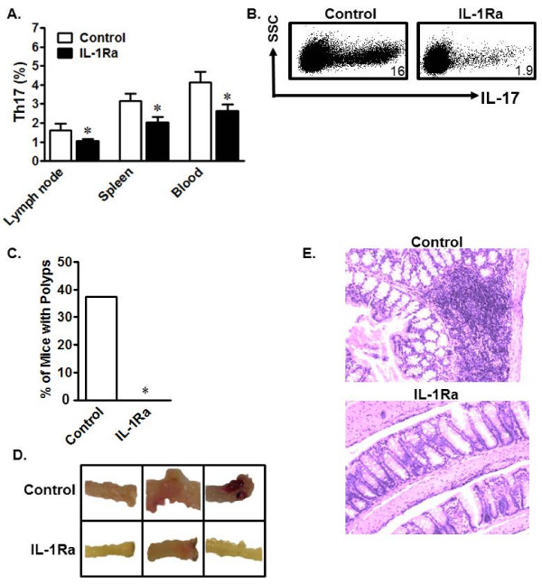
Results: We detected elevated levels of IL-17 and Th17 cells in the intestinal mucosa of patients with Crohn's disease. Intestinal DCs from Crohn's patients produced more IL-1β than controls and were superior to blood DCs in Th17 induction through an IL-1-dependent mechanism. Furthermore, IL-17 levels were negatively associated with those of IL-10 and were positively associated those of IL-1β in intestinal mucosa. These data point toward an in vivo cellular and molecular link among endogenous IL-10, IL-1, and Th17 cells in patients with Crohn's disease. We further investigated this relationship in IL-10(-/-) mice. We observed a systemic increase in Th17 cells in IL-10(-/-) mice when compared to wild-type mice. Similar to the intestinal DCs in patients with Crohn's disease, murine IL-10-/- DCs produced more IL-1β than their wild-type counterparts and promoted Th17 cell development in an IL-1-dependent manner. Finally, in vivo blockade of IL-1 receptor signaling reduced Th17 cell accumulation and inflammation in a mouse model of chemically-induced colitis.

Conclusions: Endogenous IL-10 constrains Th17 cell development through the control of IL-1 production by DCs, and reaffirms the crucial anti-inflammatory role of IL-10 in patients with chronic inflammation.


Figures

Figure 1
Figure 1
Th17 cells and IL-17 are increased in CD patient tissue. a) Lamina propria mononuclear cells from control colon tissues or CD colons were cultured for two days with IL-1 and IL-23. Supernatants were subjected to ELISA for analysis of IL-17 protein. b) Serum from healthy or CD patients was subjected to ELISA for IL-17 quantification. n = 16. *P < 0.0001. c) Healthy blood or control colon tissue or CD colon tissue cells were stained with antibodies to CD3, CD4, IL-17, and FoxP3 and antigen expression was analyzed via FACS. Results are expressed as the percent of IL-17+ or Foxp3+ T cells in the CD4+ T cells. The gates were on CD4+CD3+ T cells for both Th17 and Treg cells. n = 16.
Figure 2
Figure 2
Relationships among IL-17, IL-1, and IL-10 in CD patients. a-c) IL-17, IL-10, and IL-1β message from fresh CD mononuclear lamina propria cells were quantified via real-time PCR. Chi-squared (χ2) test. P < 0.0001 for the following comparisons: IL-17 vs IL-10 (a), IL-1β vs IL-10 (b), and IL-17 vs IL-1β (c). n = 16. d) Crohn's DCs expressed high levels of IL-1β. Crohn's DCs and macrophages, and normal colon DCs were cultured for 40 hours with or without LPS. Normal control DCs were isolated from normal colon tissues at least 5 cm away from colon cancer. IL-1β protein was quantified via ELISA. n = 6. *, P < 0.005. e) Crohn's DCs induced high levels of T cell-derived IL-17 production through IL-1. CD4+ T cells were cultured for 48 hours with Crohn's myeloid DCs or macrophages in the presence anti-IL-1R. IL-17 protein was detected via ELISA in supernatants. n = 6. *, P < 0.005.
Figure 3
Figure 3
Crohn's disease T cell-derived IL-17 stimulates the production of inflammatory cytokines. "Normal" colon tissue cells were cultured for 40 hours with medium alone or CD T cell supernatants with or without anti-IL-17R. IL-1β (a), IL-6 (b), and IL-8 (c) were quantified in supernatant. n = 5. *, P < 0.05 and **, P < 0.01.
Figure 4
Figure 4
Murine IL-10-/- DCs are powerful Th17 inducers. a, b) Increased Th17 cells in IL-10-/- mice. Mononuclear cells from unchallenged IL-10+/+ and IL-10-/- mouse organs were analyzed via FACS. a) Representative FACS plots. b) Results are expressed as the percentage of Th17 cells in CD4+ T cell populations ± SEM. 6 mice/group, *P < 0.05. c, d). IL-10-/- spleen T cells were susceptible to Th17 polarization. IL-10+/+ and IL-10-/- spleen T cells were polarized with Th17-polarizing cytokines for 6 days. Th17 cells were analyzed by FACS. c) Results are expressed as the percentage of Th17 cells in CD4+ T cells. d) Supernatant from Th17 cultures was collected on day 3 and analyzed via ELISA for levels of IL-17. *P < 0.05, average of three experiments. e) IL-10-/- DCs were potent Th17 inducers. IL-10+/+ CD4+ T cells were cultured with IL-10-/- or IL-10+/+ splenic DCs in a ratio of 5:1 for 5 days. Th17 cells were analyzed by FACS. Results are expressed as the percentage of Th17 cells in CD4+ T cells. n = 6, *P < 0.05. Y axis showed SSC and the dot plots were gated on CD4+CD3+ T cells (a, c, e).
Figure 5
Figure 5
Mouse IL-10-/- DCs induce Th17 cells through the IL-1/IL-1R signaling pathway. a) Reduced Th17 cells in DC-stimulated IL-1R-/- CD4+ T cell cultures. IL-1R-/- or IL-1R+/+ CD4+ T cells were stimulated with wild-type DCs for 5 days. The cells were analyzed for Th17. n = 3. b, c)IL-10-/- DCs expressed high levels of IL-1. IL-10+/+ or IL-10-/- DCs were cultured with or without LPS for 8 (b) or 48 hours (c). IL-1α and IL-1β message was quantified via real-time PCR (b). IL-1β protein was detected in culture supernatant via ELISA (c). *P < 0.05, average of 3 experiments. d, e) Blockade of IL-1R reduced DC-mediated Th17 induction. IL-10-/- or IL-10+/+ DCs were cultured in a ratio of 1:5 with IL-10+/+ CD4+ T cells with or without anti-IL-1R. The cells were analyzed on day 5 for Th17. Results are expressed as the percentage of Th17 cells in CD4+ T cells (d). IL-17 was detected in culture supernatants on day 3 via ELISA. Results are expressed as the mean values ± SEM (e). n = 4. *, P < 0.05. Y axis showed SSC and the dot plots were gated on CD4+CD3+ T cells (a, d).
Figure 6
Figure 6
In vivo IL-1R blockade reduces Th17 cells and inflammation. a) In vivo IL-1R blockade decreased homeostatic Th17 cells. Mice were treated with Anakinra or PBS for 7 days. Single-cell suspensions from different organs were analyzed for Th17 cells. Results are expressed as the percentage of Th17 cells in CD4+ T cells ± SEM. *P < 0.05, 5 mice/group. b) In vivo IL-1R blockade reduced DSS-induced Th17 cells. Mice were treated with DSS to induce inflammation and given Anakinra throughout the second cycle of treatment. Single-cell suspensions from different organs were analyzed for Th17 cells. Results are shown as the percentage of Th17 cells in mensenteric lymph node CD4+ T cells in mice treated with Anakinra or PBS. Y axis showed SSC and the dot plots were gated on CD4+CD3+ T cells. n = 4. c-e) In vivo IL-1R blockade prevented polyp formation and ameliorated inflammation induced by DSS. Colon polyps were counted in DSS-challenged IL-10-/- mice treated with PBS or Anakinra (IL-1Ra). n = 8 mice per group, *P < 0.05 (c). Bloody and polyp-carrying colon sections from untreated mice (top) are shown, but were not observed in IL-1Ra-treated mice (bottom) (d). H&E staining showed heavy cellular infiltration in colon tissue from untreated mice (top), but not in colon tissue from IL-1Ra-treated mice (bottom) (e).

References

    1. Tu S, Bhagat G, Cui G, Takaishi S, Kurt-Jones EA, Rickman B, Betz KS, Penz-Oesterreicher M, Bjorkdahl O, Fox JG, Wang TC. Overexpression of interleukin-1beta induces gastric inflammation and cancer and mobilizes myeloid-derived suppressor cells in mice. Cancer Cell. 2008;14:408–419. doi: 10.1016/j.ccr.2008.10.011. - DOI - PMC - PubMed
    1. Coussens LM, Werb Z. Inflammation and cancer. Nature. 2002;420:860–867. doi: 10.1038/nature01322. - DOI - PMC - PubMed
    1. Du C, Liu C, Kang J, Zhao G, Ye Z, Huang S, Li Z, Wu Z, Pei G. MicroRNA miR-326 regulates TH-17 differentiation and is associated with the pathogenesis of multiple sclerosis. Nat Immunol. 2009;10:1252–1259. doi: 10.1038/ni.1798. - DOI - PubMed
    1. Kleinschek MA, Boniface K, Sadekova S, Grein J, Murphy EE, Turner SP, Raskin L, Desai B, Faubion WA, de Waal Malefyt R. et al.Circulating and gut-resident human Th17 cells express CD161 and promote intestinal inflammation. J Exp Med. 2009;206:525–534. doi: 10.1084/jem.20081712. - DOI - PMC - PubMed
    1. Kryczek I WK, Zhao E, Wei S, Vatan L, Szeliga W, Huang E, Greenson J, Chang A, Roliński J, Radwan P, Fang J, Wang G, Zou W. IL-17+ Regulatory T Cells in the Microenvironments of Chronic Inflammation and Cancer. Journal of Immunology. 2011. - PubMed

Publication types

MeSH terms