

Sagittal plane kinematics of the adult hyoid bone
- PMID: 22176712
- PMCID: PMC3295612
- DOI: 10.1016/j.jbiomech.2011.11.040
Sagittal plane kinematics of the adult hyoid bone
Abstract
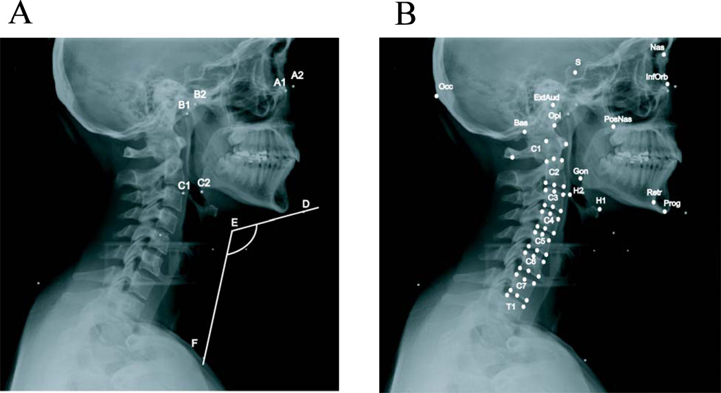
The hyoid bone is a unique bone in the skeleton not articulated to any other bone. The hyoid muscles, which attach to the hyoid bone, may play a role in neck mechanics, but analysis of their function requires quantifying hyoid bone mechanics. The goal of this study was to obtain the detailed kinematics of the hyoid bone over a large range of flexion-extension motion using radiographs at 5 postures. The position of the hyoid bone in the sagittal plane was characterized with respect to head, jaw, and vertebral movements. Sex differences in hyoid kinematics were also investigated. We hypothesized that (1) the position of the hyoid bone in the sagittal plane is linearly correlated with motion of the head, jaw, and vertebrae, and (2) the hyoid position, size, and kinematics are sex-specific. We found that the hyoid bone X, Y, and angular position generally had strong linear correlations with the positions of the head, jaw, and the cervical vertebrae C1-C4. Hyoid X and angular position was also correlated to C5. Sex differences were found in some regressions of the hyoid bone with respect to C1-C5. The angular and linear measurements of the hyoid bone showed sex differences in absolute values, which were not evident after normalization by posture or neck size. Incorporating these results to neck models would enable accurate modeling of the hyoid muscles. This may have implications for analyzing the mechanics of the cervical spine, including loads on neck structures and implants.
Copyright © 2011 Elsevier Ltd. All rights reserved.
Conflict of interest statement
None declared.
Figures


References
-
- Berzin F. Electromyographic analysis of the sternohyoid muscle and anterior belly of the digastric muscle in jaw movements. J Oral Rehabil. 1995;22:463–467. - PubMed
-
- Bibby RE, Preston CB. The hyoid triangle. Am J Orthod. 1981;80:92–97. - PubMed
-
- Brodie AG. Anatomy and physiology of head and neck musculature. Am J Orthod. 1950;36:831–844. - PubMed
-
- Chancey VC, Nightingale RW, Van Ee CA, Knaub KE, Myers BS. Improved estimation of human neck tensile tolerance: reducing the range of reported tolerance using anthropometrically correct muscles and optimized physiologic initial conditions. Stapp Car Crash J. 2003;47:135–153. - PubMed
-
- Crompton AW, Cook P, Hiiemae K, Thexton AJ. Movement of the hyoid apparatus during chewing. Nature. 1975;258:69–70. - PubMed
Publication types
MeSH terms
Grants and funding
LinkOut - more resources
Full Text Sources
Miscellaneous
