. 2012 Mar;12(3):576-89.
doi: 10.1111/j.1600-6143.2011.03879.x. Epub 2011 Dec 17.

Fates of CD4+ T cells in a tolerant environment depend on timing and place of antigen exposure

Affiliations

Fates of CD4+ T cells in a tolerant environment depend on timing and place of antigen exposure

B E Burrell et al. Am J Transplant. 2012 Mar.

Abstract

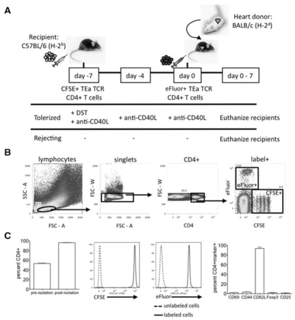
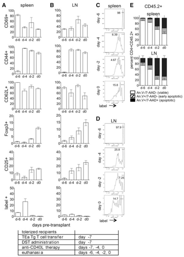
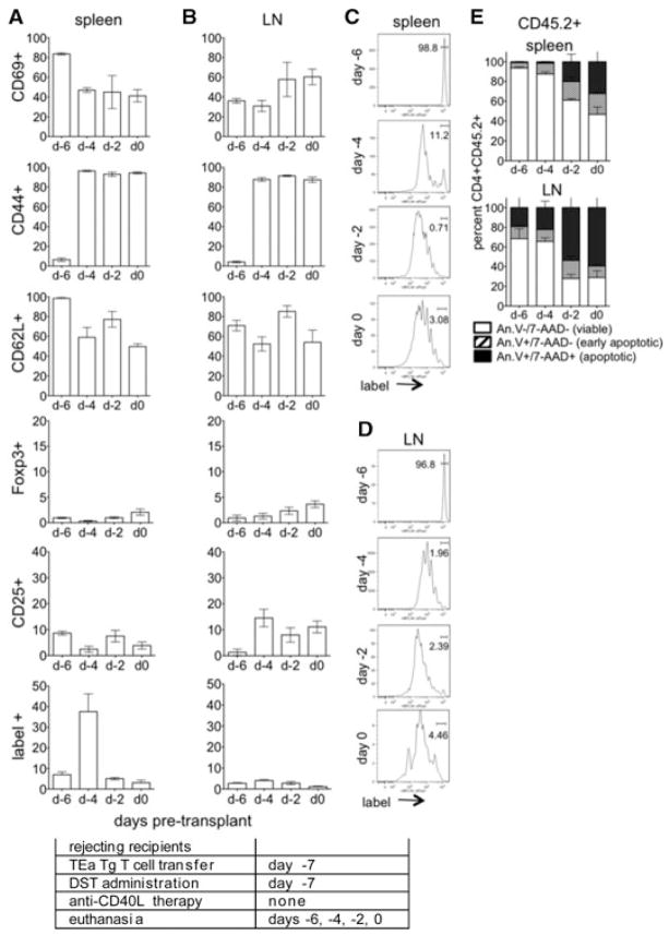
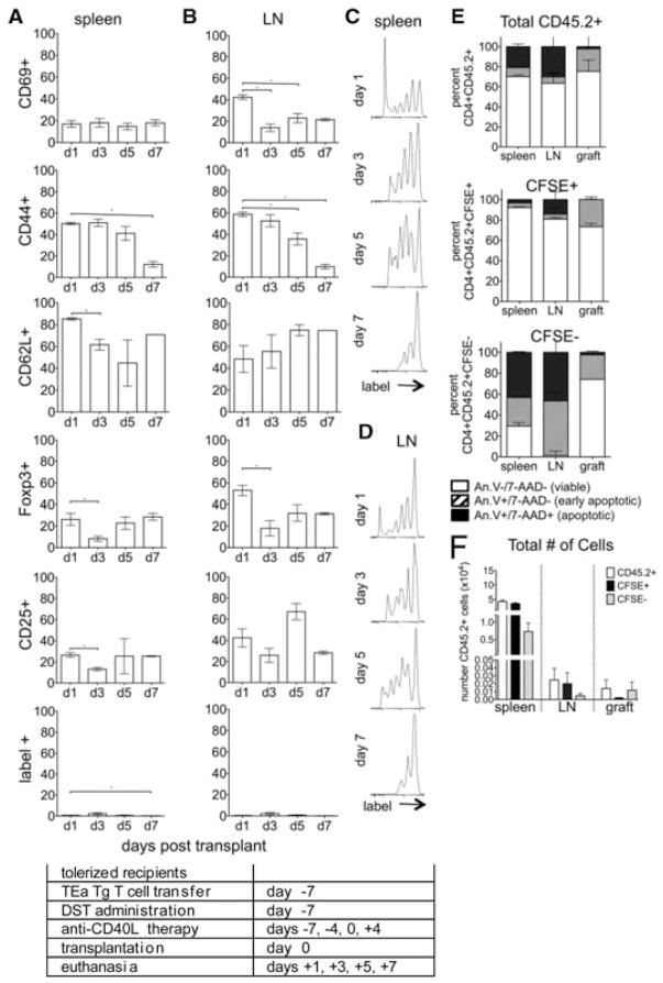
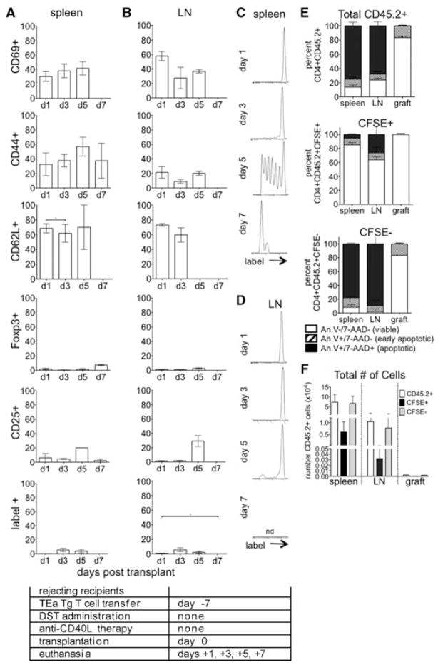
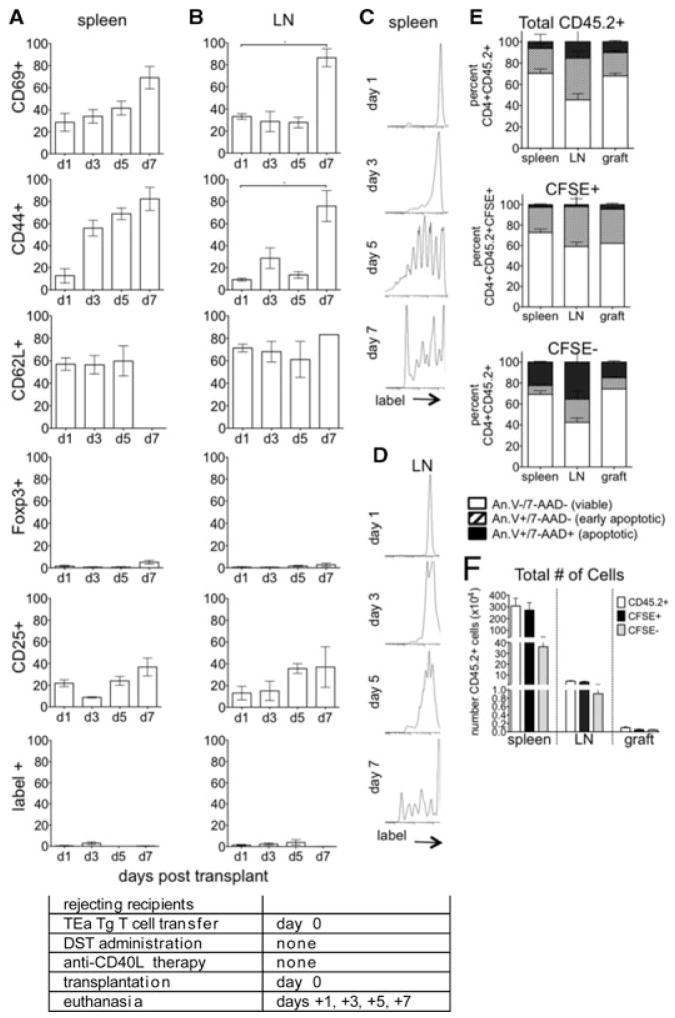
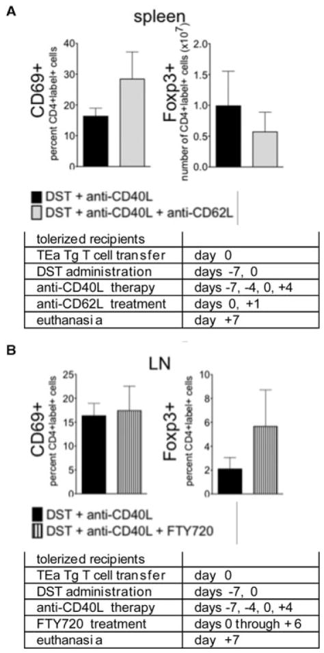
In experimental organ transplantation, tolerance is induced by administration of anti-CD40L mAb in conjunction with donor-specific splenocyte transfusion. Multiple, sometimes conflicting mechanisms of action resulting from this treatment have been reported. To resolve these issues, this study assessed the fates of graft reactive cells at different times and locations in the tolerant environment. Alloantigen-specific CD4(+) T cells transferred at time of tolerance induction (7 days before transplantation) became activated, expressed CD69 and CD44, and proliferated. Importantly, a large subset of this population became Foxp3(+) , more so in the lymph nodes than spleen, indicative of differentiation to a regulatory phenotype. In contrast, graft reactive CD4(+) T cells transferred to tolerogen-treated recipients at the time of transplantation failed either to proliferate or to differentiate, and instead were deleted via apoptosis. In untreated rejecting recipients graft reactive CD4(+) T cells became activated, proliferated and differentiated mainly in the spleen, and many of these cells were eventually deleted. These data resolve many apparent contradictions in the literature by showing that the timing of antigen exposure, the immunologic status of the recipients and secondary lymphoid organ location act together as key factors to determine the fate of graft reactive CD4(+) T cells.


Conflict of interest statement

Disclosure

The authors of this manuscript have no conflicts of interest to disclose as described by the American Journal of Transplantation.

Figures

Figure 1
Figure 1. Isolation and transfer of naive alloantigen-specific CD4+ T cells to track the fate of alloreactive cells in cardiac al-lograft recipients
(A) C57BL/6 recipients received 2 × 10⁷ CFSE-labeled TEa TCR Tg CD4+ cells 7 days before transplant (day –7). On the day of transplantation (day 0), recipients received 2 × 10⁷ eFluor-labeled TEa TCR Tg CD4+ T cells along with a BALB/c cardiac allograft. Tolerized mice were given 107 BALB/c splenocytes (DST) i.v. on day –7 and 250 μg anti-CD40L mAb on days –7, –4, 0 and +4 relative to transplant. Rejecting control mice were left untreated. Recipients were euthanized 0–7 days posttrans-plant. (B) Gating scheme. Flow cytometric dot plots were gated on label+CD4+ singlet lymphocytes. Representative staining of splenic lymphocytes is depicted. (C) Purity, labeling and phenotype of cells pre-transfer. TEa TCR Tg CD4+ T cells were enriched from the spleen and LN of TEa mice as described in Materials and Methods. Percentage of cells that were CD4+ before and after isolation (left panel) and representative relative fluorescence histograms after labeling with CFSE (middle left panel) and eFluor (middle right panel) are shown (n = 28). The naive phenotype of transferred cells was confirmed by the presence or absence, as appropriate, of CD69, CD44, CD62L, Foxp3 and CD25 (right panel, n = 3).
Figure 2
Figure 2. Graft reactive cells proliferate and become activated in response to treatment with DST and anti-CD40L mAb before transplantation
(A–D) CD4+ TEa cells (CD45.2+) were isolated, labeled with eFluor and transferred to tolerogen (DST + anti-CD40L mAb) treated congenic CD45.1+ C57BL/6 mice at day –7. Recipients were euthanized on days –6, –4, –2 and 0, and CD45.2+CD4+label+ splenocytes (A, C) and LN lymphocytes (B, D) analyzed for CD69, CD44, CD62L, Foxp3 and CD25 and for proliferation by label dilution. (E) For apoptosis, total transferred TEa cells (CD45.2+) were recovered from the spleen and LN and analyzed by annexin V and 7-AAD staining on days –6, –4, –2 and 0. Data represent of 4 mice/group. Experimental conditions are outlined in the accompanying table.
Figure 3
Figure 3. Graft reactive cells proliferate and become activated in response to treatment with DST alone
(A–D) CD4+ TEa cells (CD45.2+) were isolated, labeled with eFluor and transferred to DST-treated congenic CD45.1+ C57BL/6 mice at day –7. Recipients were euthanized on days –6, –4, –2, and 0 and CD45.2+CD4+label+ splenocytes (A, C) and LN lymphocytes (B, D) analyzed for CD69, CD44, CD62L, Foxp3 and CD25, and for proliferation by label dilution. (E) For apoptosis, total transferred TEa cells (CD45.2+) were recovered from the spleen and LN and analyzed by annexin V and 7-AAD staining on days –6, –4, –2 and 0. Data represent of 4 mice/group. Experimental conditions are outlined in the accompanying table.
Figure 4
Figure 4. Graft reactive cells transferred at day –7 undergo phenotypic changes in response to tolerance induction
(A–D) CD4+ TEa cells were isolated, labeled with CSFE and transferred to tolerogen (DST and anti-CD40L mAb) treated C57BL/6 allograft recipients at day –7 relative to transplant. Recipients were euthanized on days 1, 3, 5 and 7 and CD4+label+ splenocytes (A, C) and LN lymphocytes (B, D) analyzed for CD69, CD44, CD62L, Foxp3 and CD25 and for proliferation by label dilution. (E) For apoptosis, congenic recipients (CD45.1+) received TEa cells (CD45.2+) on day –7 and were euthanized day 1 posttransplantation, day 8 postexperimental onset. Total transferred TEa cells (CD45.2+, top), TEa cells that maintained their label (CFSE+, middle), and TEa cells that proliferated sufficiently to lose their label (CFSE, bottom) were recovered from the spleen, LN and graft and analyzed by annexin V and 7-AAD staining. (F) The total number of CD45.2+, CFSE+ and CFSE cells recovered from the spleens, LN and grafts and analyzed in (E) are represented. p < 0.05. Data represent of 3–8 transplanted mice/group. Experimental conditions are outlined in the accompanying table.
Figure 5
Figure 5. Graft reactive cells exposed to antigen in a tolerant environment do not become activated
eFluor labeled CD4+ TEa cells were transferred to tolerized recipients on the day of transplantation (day 0). Recipients were euthanized at days 1, 3, 5 and 7 and CD4+label+ splenocytes (A, C) and LN lymphocytes (B, D) analyzed for (A, B) ac- tivation and Treg conversion, (C, D) proliferation and (E) apoptosis as in Figure 3. (F) The total number of CD45.2+, CFSE+ and CFSE cells recovered from the spleens, LN and grafts (5 days post-day 0 cell transfer, 13 days postexperimental onset) and analyzed in (E) are represented. p < 0.05. Data represent of 3–8 transplanted mice/group. Experimental conditions are outlined in the accompanying table.
Figure 6
Figure 6. Graft reactive cells transferred at day –7 undergo phenotypic changes in response to rejection
CFSE labeled CD4+ TEa cells were transferred to untreated allograft recipients at day –7. Recipients were euthanized at days 1, 3, 5 and 7 and CD4+label+ splenocytes (A, C) and LN lymphocytes (B, D) analyzed for (A, B) activation and Treg conversion, (C, D) proliferation and (E) apoptosis as in Figure 3. (F) The total number of CD45.2+, CFSE+ and CFSE cells recovered from the spleens, LN and grafts (8 days pos-texperimental onset) and analyzed in (E) are represented. nd, insufficient cell number to provide accurate proliferative data; p < 0.05. Data represent of 3–8 transplanted mice/group. Experimental conditions are outlined in the accompanying table.
Figure 7
Figure 7. Graft reactive cells transferred at the time of transplantation undergo phenotypic changes in response to rejection
eFluor labeled CD4+ TEa cells were transferred to untreated recipients on the day of transplantation (day 0). Recipients were euthanized at days 1, 3, 5 and 7 and CD4+label+ splenocytes (A, C) and LN lymphocytes (B, D) analyzed for (A, B) activation and Treg conversion, (C, D) proliferation and (E) apoptosis as in Figure 3. (F) The total number of CD45.2+, CFSE+ and CFSE cells recovered from the spleens, LN and grafts (5 days post-day 0 cell transfer, 13 days postexperimental onset) and analyzed in (E) are represented. p < 0.05. Data represent of 3–8 transplanted mice/group. Experimental conditions are outlined in the accompanying table.
Figure 8
Figure 8. Lymph node occupancy does not guarantee activation or Foxp3 expression by graft reactive cells transferred at day 0
Tolerance was induced on day –7, and on day 0 mice received another dose of DST and transfer of eFluor labeled TEa cells. Recipients were euthanized 7 days after the second DST, 14 days after the onset of the experiment, and cells transferred on day 0 found in the spleen and LN were analyzed for expression of CD69 and Foxp3. Recipients additionally received anti-CD62L mAb on days 0 and 1 (100 μg/dose, gray bars) or FTY720 daily from day 0–6 (25 μg/dose, striped bars). Data are represented as the total number or percentage of the indicated cells recovered from spleen or LN. n = 5 mice for each condition, p > 0.05 for all data sets. Experimental conditions are outlined in the accompanying tables.

Comment in

  • Timing is everything in tolerance.
    Burlingham WJ. Burlingham WJ. Am J Transplant. 2012 Mar;12(3):517-8. doi: 10.1111/j.1600-6143.2011.03877.x. Epub 2011 Dec 17. Am J Transplant. 2012. PMID: 22176723 Free PMC article. No abstract available.

References

    1. Zheng XX, Sanchez-Fueyo A, Domenig C, Strom TB. The balance of deletion and regulation in allograft tolerance. Immunol Rev. 2003;196:75–84. - PubMed
    1. Rothstein DM, Sayegh MH. T-cell costimulatory pathways in allograft rejection and tolerance. Immunol Rev. 2003;196:85–108. - PubMed
    1. Halloran PF. Immunosuppressive drugs for kidney transplantation. N Engl J Med. 2004;351:2715–2729. - PubMed
    1. Knechtle SJ, Hamawy MM, Hu H, Fechner JH, Jr, Cho CS. Tolerance and near-tolerance strategies in monkeys and their application to human renal transplantation. Immunol Rev. 2001;183:205–213. - PubMed
    1. Durrbach A, Francois H, Jacquet A, Beaudreuil S, Charpentier B. Co-signals in organ transplantation. Curr Opin Organ Transplant. 2010;15:474–480. - PubMed

Publication types

MeSH terms