

Role of mucus layers in gut infection and inflammation
- PMID: 22177113
- PMCID: PMC3716454
- DOI: 10.1016/j.mib.2011.11.002
Role of mucus layers in gut infection and inflammation
Abstract
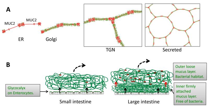
The intestinal mucus is an efficient system for protecting the epithelium from bacteria by promoting their clearance and separating them from the epithelial cells, thereby inhibiting inflammation and infection. The function of the colon inner mucus layer is especially important as this explains how we can harbor the large number of bacteria in our gut. The major component of this mucus system is the MUC2 mucin which organizes the mucus by its enormously large net-like polymers. Pathogenic microorganisms, in turn, have developed mechanisms for circumventing this well-organized mucus protective system.
Copyright © 2011 Elsevier Ltd. All rights reserved.
Figures
References
-
-
Lang T, Hansson GC, Samuelsson T. Gel-forming mucins appeared early in metazoan evolution. Proc.Natl.Acad.Sci.USA. 2007;104:16209–16214. Describes the evolution of mucins and especially that the typical domain arrangement in the gelforming mucins is found already among metazoans.
-
-
-
Atuma C, Strugula V, Allen A, Holm L. The adherent gastrointestinal mucus gel layer: thickness and physical state in vivo. Am.J.Physiol. 2001;280:G922–G929. The mucus thickness throughout the gastrointestinal tract was measured on live, anaestesized rats.
-
-
-
Johansson MEV, Phillipson M, Petersson J, Holm L, Velcich A, Hansson GC. The inner of the two Muc2 mucin dependent mucus layers in colon is devoid of bacteria. Proc.Natl.Acad.Sci.USA. 2008;105:15064–15069. This show that the inner mucus layer is separating the colon bacteria from the epithelial cells. It also shows that lack of this mucus layer allow bacteria to contact the epithelia, enter the crypts and the the epithelial cells, something that triggers inflammation.
-
Publication types
MeSH terms
Grants and funding
LinkOut - more resources
Full Text Sources
Other Literature Sources
Miscellaneous
